|
The patient came
to
the clinic 20-November-2006 complaining of left sciatica with
LBP for 3 years. Exacerbation the last 5 months down to S1
territory. MRI lumbar spine performed 14-September-2006 showed bulge
disc L3-4 with
Tarlov cyst in the sacral area. The patient has
in anamnesis
infective endocarditis with anterior mitral leaflet
prolapse. The patient is Yemeni citizen and he was operated at
home 01-November-2006. Excision of Tarlov cyst was performed, after
what a huge subcutaneous CSF pocket took place, with headache upon
movement, setting and standing.
On examination: the patient has weak left upper limb with
hypalgesia of the median nerve distribution. He had hypalgesia of
the left S1 root territory. The collection was not oozing and the
silk sutures still in place and compression of the collection caused
headache.
MRI of the lumbar spine with contrast with MR Myelography was
performed 21-November-2006 which confirmed the presence of huge CSF
collection.
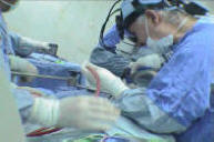
2 gm of Rocephine were given with gentamycin 80 mg before the
operation. The old incision was opened with the patient in supine
and Reverse Trendelenburg position, so that the sacral area was the upper
most. Inspection of the wound revealed a dural defect in the right
S3 root about 2 mm in length. Using Nylon 6 zero the dural
defect was sutured water-tightly, trying not to touch the running
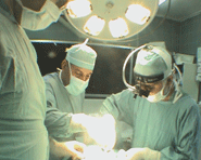
adherent root inside the dural sleeve. The patient was
positioned so that the head is in high position and Valsalva
maneuver was performed to check for CSF leak. No CSF leak. The
running roots were covered with surgicele and above that chips of
bone harvested during exposure were implanted above the sacral
defect for future bone formation to close the bone defect.
Multilayer water-tight closure of the wound and the skin was trimmed
for cosmetic reasons and subcuticular closure performed. Smooth
postoperative recovery.
Comments:
1. In our practice, we usually operate upon Tarlov cysts, when they
grow over protracted period of time. Many cases do not need surgery
and they stay under observation. Considering that, it was not
understandable the indications for such surgery, from the first
visit.
2. When surgery was needed for growing Tarlov cysts, during the last
27 years, I usually used the bipolar to shrink the cyst wall, so
that the cyst disappear completely without opening it and the root
regain normal anatomy. After that, the cavity is filled with fat
tissue. Using this technique, there were no such complications in
the past.
3. The patient still undiagnosed up to now. When the complication
resolve, the patient needs further investigations to know the cause
of his complains. |