Although the nerves of the upper extremity can be studied in a variety of ways, the approach taken here focuses on regional anatomy. The nerves of the upper extremity have therefore been divided into three regions of surgical exposure:
1. the subaxilla and upper arm (which include the nerves of the distal brachial plexus and the musculocutaneous, median ulnar, radial, and axillary nerves)
2. the elbow and forearm (which include the median, ulnar, and radial nerves on the anterior and medial aspects of the elbow and forearm)
3. the palm and wrist (which include the median and ulnar nerves on the anterior aspect of the wrist and palm, as well as the digital nerves)
Variants of the nerves of the upper extremity are presented. The implications of these variants, both clinical and surgical, are addressed.
Subaxilla and Upper Arm
Lower Brachial Plexus
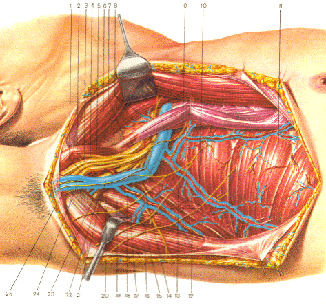
The lower brachial plexus is approached through an infraclavicular brachial plexus incision, that extends over the pectoralis muscles' insertion on the humerus and drops down onto the intermuscular septum (between the biceps and the triceps muscles). The incision of the pectoralis major muscle should be made in close proximity to the humerus while leaving enough tendon for suturing at the time of closure. If the incision is made too far from the humerus, the tendon thins and is inadequate for a strong suture fixation to the insertion site. Following the reflection of the pectoralis major muscle medially (if indicated), the cords of the brachial plexus and the proximal nerves of the upper arm are easily observed and identified. In this region, the relationship of the axillary (brachial) artery to the radial, median, and ulnar nerves is consistent and aids the surgeon in nerve identification.
| Fig-1: Topographical presentation of the subaxillary region
1- n. ulnaris; 2- n. cutaneous antebrachii medialis; 3- n. medianus; 4- n. musculocutaneous; 5- m. coracobrachialis; 6- m. pectoralis major; 7- m. pectoralis minor; 8- rami pectorales a. thoracoacrommialis; 9- nn. thoracales anteriores; 10- a & v. thoracica lateralis and n. thoracicus longus ; 11- m. serratus anterior; 12- a. & v. thoracodorsalis; 13- nn. intercostobrachialies; 14- n. thoracodorsalis; 15- a. & v. circumflexa scapule; 16- m. latissimus dorsi & m. teres major; 17-foramen trilaterum; 18- a. & v. subscapularis; 19- m. subscapularis; 20- caput longum m tricipitis brachii; 21- v. axillaris; 22- foramen quadrilaterum; 23- n. axillaris; 24- n. cutaneus brachii medialis; 25- n. radialis.
Proximal Brachial Nerves
The proximal axillary and musculocutaneous nerves are readily visualized with this approach. The brachial and antebrachial cutaneous nerves also may be identified in the intermuscular septum. It is important to recognize their proximal separation from their parent nerve (ulnar nerve). The antebrachial cutaneous nerve does not pass through the cubital fossa with the ulnar nerve despite its parallel relationship over a significant portion of its course. The thoracodorsal nerve is visualized along the chest wall. This nerve readily is observed following medial chest wall exposure. Finally, the radial nerve (a direct extension of the posterior cord) is visualized just before it passes behind the humerus as it begins its descent by spiraling around the humerus.
Medial Upper Arm
The musculocutaneous, median, and ulnar nerves are located in the medial aspect of the upper arm. Both the median and ulnar nerves descend in the intermuscular septum, along the brachial artery and vein. Their relationship to these vascular structures as well as their relationship to the medial brachial and antebrachial cutaneous nerves are illustrated in Figures 1. The medial arm incision described before gains access to the intermuscular septum as described. It is emphasized that this region is straightforward with respect to anatomic relationships; therefore, an exhaustive discussion of the anatomic details will not be undertaken. The musculocutaneous nerve is readily visualized by retracting the biceps muscle. Its branches, which innervate the biceps and brachialis muscles, can then be observed.
Lateral and Posterior Upper Arm
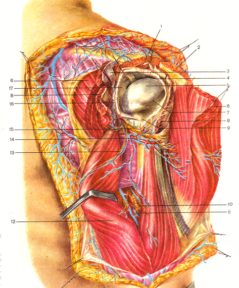
In contrast to the medial upper arm, the lateral and posterior upper arm is more complex regarding peripheral nerve exposure. As the radial nerve spirals around the humerus, it passes under and through several muscles enroute to the forearm. An incision, over the medial aspect of forearm, gains access to the entire course of the radial nerve in the proximal upper extremity, in addition to the distal axillary nerve (the proximal axillary nerve may be approached best through the subaxillary approach). Following opening of the brachial fascia (which is a fairly significant fascial covering), the axillary nerve as it approaches the deltoid muscle is observed. The separation of the triceps muscle bellies (long and lateral heads) along their fibers allows a view and exposure of the radial nerve throughout its course in the upper arm. This is achieved by simply mobilizing the muscle bellies from side to side. After mobilizing these muscles, the radial nerve can be observed underneath. The planes between the muscle bellies can be identified by palpation. Blunt dissection allows their separation and, thus, the desired deeper exposure (Figure 2). A radial compressive neuropathy caused by the triceps muscle in this region may occur. The radial nerve spirals around the humerus, staying in relatively close proximity to the bone. Distally in the upper arm, however, it pierces an intermuscular septum between the brachioradialis and the brachialis muscles. Reflection of the brachioradialis muscle laterally allows the reidentification of the nerve medial to the muscle and underneath the septum. Care must be taken to minimize lateral muscle dissection, which could injure the motor branches to the muscle. Multiple motor branches and the posterior antebrachial cutaneous nerve branch from the radial nerve and can be visualized through this exposure.
Elbow and Forearm Region
Ulnar Nerve
The cubital tunnel begins at the condylar groove between the medial epicondyle of the humerus and the olecranon of the ulna. The floor of the cubital tunnel is the medial collateral ligament (ulnar lateral ligament) of the elbow joint, and the sides are formed by the two heads of the flexor carpi ulnaris muscle. The roof is formed by the triangular arcuate ligament (aponeurotic band) that bridges from the medial epicondyle of the humerus to the medial aspect of the olecranon. The capacity of the cubital tunnel is greatest when the elbow is in extension. Measurements in cadaveric material demonstrate that the distance between the humeral and ulnar attachments of the triangular arcuate ligament lengthens 5 mm for each 45 degrees of flexion. After penetrating the flexor carpi ulnaris muscle, the ulnar nerve continues throughout the entire forearm under the cover of the flexor carpi ulnaris muscle (Figure 3). Just above the middle of the forearm, the ulnar artery veers toward the nerve from the radial aspect and continues to lie close to the nerve until it reaches the wrist joint. The neurovascular bundle becomes more superficial in the distal third of the forearm, where it is barely under cover of the radial border of the flexor carpi ulnaris muscle. The surgical approach to the ulnar nerve at the elbow is through a posteromedial longitudinal curvilinear or zigzag incision. The ulnar nerve should be freed for at least 8 cm proximal to the medial epicondyle to ensure release from the arcade of Struthers or entrapment on the medial intermuscular septum. Fixation of the nerve at the level of the medial epicondylar groove can result in traction neuritis. The fibrous arcade over the flexor carpi ulnaris muscle must be excised, and the ulnar nerve should be explored to the midportion of the proximal third of the forearm. The superior ulnar collateral artery, which accompanies the ulnar nerve in the condylar groove, should not be injured. Exposure of the ulnar nerve in the forearm is best accomplished through a longitudinal "stepped" or 60-degree "turned" incision along the ulnar border of the forearm about 5 cm volar to the palpable portion of the ulnar bone. After opening the antebrachial fascia, the nerve is approached by separating the flexor carpi ulnaris from the flexor digitorum superficialis muscle. After creating this division, the flexor carpi ulnaris is elevated from the flexor digitorum profundus to expose the ulnar nerve throughout the forearm. The cubital tunnel syndrome and, occasionally, median nerve entrapment can occur in the region of the elbow. Nerve injury from percutaneous or open attempts at vascular access or angiography may result in median nerve injury as well.
Median Nerve
The median nerve begins just below the pectoralis minor muscle as a union of branches from the lateral and medial cords of the brachial plexus. The median nerve continues on the medial side of the biceps brachii muscle and accompanies the brachial artery to the elbow. The median nerve is closely related to the vessels through most of the arm, and in those injuries that damage the vessel, the nerve is almost always involved. The nerve lies in the medial bicipital groove and anterior to the medial intermuscular septum. As it approaches the elbow joint, the median nerve is directly covered by the lacertus fibrosis (bicipital aponeurosis)(Figure-4). The nerve passes out of the cubital fossa between the humeral and ulnar heads of the pronator teres muscle. Just beyond the exit of the median nerve from between the heads of the pronator teres, the anterior interosseous nerve branch passes forward to supply the deep muscles of the forearm. The median nerve passes behind the flexor superficialis arch and comes to lie deeply in the middle of the volar surface of the forearm, between the flexor digitorum superficialis and the flexor digitorum profundus muscles. The median nerve becomes superficial to the flexor digitorum superficialis muscle bellies about 5 cm proximal to the transverse carpal ligament. The operative approach to the median nerve in the forearm is a longitudinal approach along the medial ulnar border of the biceps brachii muscle in the distal arm, curving transversely at the elbow flexion crease, then taking either a gentle S-curve or a zigzag course along the ulnar (medial) distal border of the pronator teres muscles, and continuing longitudinally to the wrist crease. An incision into the palm should cross the wrist crease obliquely at a point in line with the long axis of the ring finger. The safe side of the median nerve in the proximal forearm is the radial side, since the branches are ulnarward. The best surgical approach is to identify the median nerve proximal to the lacertus fibrosis and to trace it distally through the region of the pronator teres and the flexor superficialis arch. Entrapment of the median nerve in the proximal forearm is termed the pronator syndrome. Most commonly the entrapment occurs by the deep head of the pronator muscle. At least two other areas of potential entrapment exist in close proximity: at the lacertus fibrosis and at the flexor superficialis arch. When forearm pain is reproduced by resistance to pronation of the forearm, and is aggravated by extending the elbow, the localization is to the pronator teres. When the pain is reproduced by resistance to flexion of the elbow and supination of the forearm, the lacertus fibrosis is the likely site of compression. Pain in the proximal forearm, reproduced by resistance to flexion of the flexor digitorum superficialis of the long finger, helps to localize the pathology to the flexor superficialis arch. When surgical release of a pronator syndrome is necessary, the median nerve is first identified proximal to the lacertus fibrosis and then followed distally through the flexor superficialis bridge. It is necessary to develop the plane between the lower border of the pronator teres and the proximal margin of the flexor carpi radialis. The anterior interosseous nerve syndrome is a result of compression of the anterior interosseous branch of the median nerve, usually at a site close to its branching. The anterior interosseous nerve usually arises from the median nerve approximately 7 cm distal to the lateral epicondyle. The anterior interosseous nerve syndrome is characterized by an inability to flex the terminal phalanges of the thumb, index, and long fingers. Sensation in the hand is undisturbed. The median nerve should be identified proximal to the lacertus fibrosis and traced distally through the pronator teres. The most common restraining structure is the tendinous origin of the deep head of the pronator teres.
Combined Surgical Exposure of the Median and Ulnar Nerves
The exposure of the median and ulnar nerves in the region of the elbow may be performed through one incision by simply carrying the medial upper arm incision caudally in an S-shaped fashion. In the region of the elbow, the single incision approach is not often used because a more limited exposure to just one of the nerves is indicated. A limited exposure is used when appropriate. At the level of the elbow, the single incision gains access to the antecubital fossae and the cubital tunnel where the median and ulnar nerves, respectively, pass. Exposure of both of these nerves is straightforward in this region. If any difficulty is encountered in locating either of these nerves during a surgical exposure due to anatomic variation or scarring from previous injury or surgery, it would be prudent to find the nerve by a more aggressive and more proximal exposure. Approximately 8 cm proximal to the medial epicondyle, the ulnar nerve normally passes from the anterior plane of the medial intermuscular septum to the posterior plane of the septum. The arcade of Struthers is formed by a thickening of the deep investing fascia of the distal part of the arm, by superficial muscular fibers of the medial head of the triceps, and by attachments of the internal brachial ligament. Its anterior border is the medial intermuscular septum. Proximal to the elbow, the ulnar nerve should be freed for at least 8 cm from the medial epicondyle to avoid potential compression in the arcade of Struthers. If both the median and ulnar nerves require exposure in the distal forearm, a single incision can be used. Several authors recommend a longitudinal incision midway between the two nerves. This should be an "S-course" with angles 90 degrees or more. The antebrachial fascia is opened and the interval developed between the palmaris longus and flexor carpi ulnaris muscles. The ulnar nerve throughout the forearm is in the plane between the flexor carpi ulnaris and the flexor digitorum profundus muscles. The median nerve lies beneath the flexor digitorum superficialis. This incision can provide good exposure of both nerves in the distal two-thirds of the forearm. |
|
| 
|
|
|
Fig-2: Topographical view of the shoulder from behind.
1- m. supraspinatus; 2- acromion; 3- m. infraspinatus; 4- tendon of the long head of m. bicipitis brachii; 5- caput humeri; 6- deltoid muscle; 7- collum anatomicum; 8- m. teres minor; 9- collum chirurgicum; 10- n. radialis; 11- collateral radial artery; 12- caput longum m tricipitis brachii; 13- n. cutaneus brachii lateralis; 14- a. circumflexa humeri posterior; 15- n. axillaris; 16- capsula articularis; 17- labrium glenoidale. |
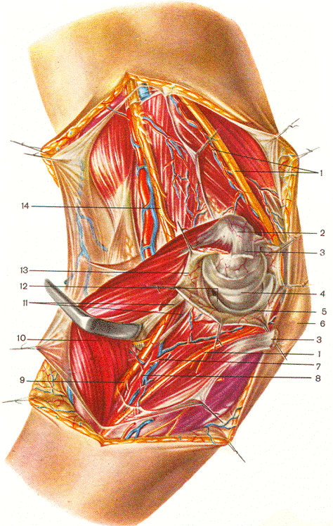 |
Fig-3: Topography of the elbow - Volar view
1- ulnar nerve with it's collateral artery; 2- medial epicondyle; 3-m.flexor carpi ulnaris; 4- trochlea humeri; 5- incissura trochlearis ulnae; 6- olecranon; 7- a. recurrence ulnaris; 8- ulnar a.; 9- median n.; 10- m.flexor digitorum superficialis; 11- pronator teres m. 12- processus coronoideus ulnae; 13- capsula articularis of elbow joint; 14- median n. |
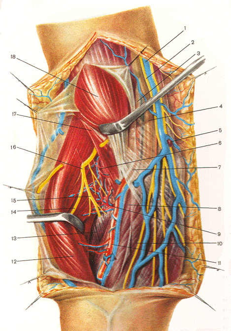
Fig-4: Anatomical relations of the neurovascular bundles in the elbow.
1- v. basilica; 2- n. cutaneus antebrachii medialis; 3- a. brachialis; 4- median n.; 5- lymphatic node; 6- radial collateral a.; 7- aponeurosis brachial biceps m.; 8- pronator teres m.; 9- recurrent radial a.; 10- flexor carpi radialis m.; 11- radial a.; 12- m. brachioradialis; 13- supinator m.; 14- superficial branch of radial n.; 15- deep branch of radial n.; 16- radial n.; 17- brachial m.; 18- m. biceps brachii. |
Fig-5: Topography of the elbow -lateral view.
1. humerus; 2- septum intermuscularis brachii lateralis; 3-caput laterale m. tricipitis brachii; 4- m. bachioradialis; 5- m. extensor carpi radialis longus; 6- superficial branch of the radial n.; 7- extensor carpi radialis brevis; 8- extensor digitorum; 9- supinator; 10- deep branch of the radial n.; 11- collum radii; 12- caput radii; 13- incisura trochlearis ulnae; 14- capitulum humeri; 15- trochlea humeri; 16- lateral epicondyle. |
Radial Nerve
The radial nerve, as it courses across the region of the elbow, follows a complex anatomic course. Reflection of the brachioradialis and the extensor radialis longus muscles allows the exposure of the radial nerve as it crosses the elbow and maintains its submuscular descent into the forearm. In the region of the elbow, it branches into a superficial and deep branch. The superficial branch descends into the forearm between and underneath the brachioradialis and extensor carpi radialis longus muscles. The deep branch (the posterior interosseous nerve) passes underneath the flexor carpi radialis longus muscle. It is emphasized again that with careful dissection and retraction of the muscles under which the radial nerve passes, it can be safely followed (without significant nervous or other soft-tissue injury) over its entire course into the forearm. Protection of the motor branches and an understanding of the location of the complicated regions of its exposure are critical. Exposure of the radial nerve as it passes distally toward the elbow is performed with an incision along the medial border of the brachioradialis muscle (Figure -4). Reflection of the brachioradialis muscle laterally allows the reidentification of the radial nerve. Care must be taken to minimize lateral muscle dissection in this segment, which could injure motor branches to the muscle. Multiple motor branches, and the posterior antebrachial cutaneous nerve, branch from the radial nerve in this segment and can be visualized through this exposure. The radial nerve as it passes across the region of the elbow is a similarly complex anatomic exposure. An anterolateral incision should extend from 8-10 cm proximal to the biepicondylar line and end about 5-6 cm distal to the line. Lateral reflection of the brachioradialis and the extensor radialis longus muscle allows the exposure of the radial nerve as it crosses the elbow and maintains its submuscular descent into the forearm. The biceps tendon and muscle, together with the musculocutaneous nerve, are retracted medially. The musculocutaneous nerve lies at more superficial level than the radial nerve. In the region of the elbow, the radial nerve branches into a superficial sensory branch and a deep motor branch, usually at the level of th radiocapitellar joint, but in a range of 5 cm proximal or distal to this point. The superficial branch descends into the forearm between and underneath the brachioradialis and extensor carpi radialis muscles. The superficial radial nerve is a key to accurate exposure. It is trace proximally to the radial nerve and then distally to the posterior interosseous nerve. The deep branch, the posterior interosseous nerve, passes underneath the flexor carpi radialis longus muscle and between the two heads of the supinator muscle. The nerve lies deep in the cubital fossa. The incision should be generous in order to obtain adequate visualization. Common syndromes with radial nerve injury include one associated with the compression of the radial nerve as it passes through the intermuscular septum, "Saturday night palsy" (caused by prolonged pressure exerted on the nerve as it passes around the proximal humerus), fracture of the proximal humerus (associated with injury to the closely approximated radial nerve), and injury to the nerve in the region of the radial tunnel (which comprises the tunnel under and around the brachioradialis and extensor carpi radialis longus muscles, the capitulum, the biceps tendon, the head of the radius, the pronator teres, the extensor carpi radialis brevis, and the superficial and deep heads of the supinator muscles in descending order). The latter region has been loosely referred to as the radial tunnel, and the syndrome associated with it as the resistant tennis elbow syndrome. The superficial radial nerve in the forearm continues in its position beneath the extensor carpi radialis longus muscle to the junction of the middle and lower thirds of the forearm. It pierces the deep fascia and supplies cutaneous branches to the radial posterior aspect of the carpus, and the thumb, index, and middle fingers. The point of superficial penetration is 8-9 cm proximal to the tip of the radial styloid process, or just above the junction of the middle and lower thirds of the forearm. This nerve can be exposed by a longitudinal incision on the radiodorsal aspect of the distal upper arm or anteriorly on the proximal forearm. The most serious complication of elective surgery in this area is a painful neuroma of one of the branches of the superficial radial nerve, which may follow laceration, retraction stretch, or blunt injury, and is termed cheiralgia paresthetica. This region is exposed to everyday activity-related trauma, while wrist movement is a further irritant. Therefore, multiple factors serve to prolong discomfort. The radial nerve and its major branches at the elbow can be exposed in the lower arm and forearm through an incision that begins anteriorly between the brachialis and the brachioradialis muscles and extends dorsally into the forearm between the extensor carpi radialis brevis and the extensor digitorum communis muscle. The plane between the extensor carpi radialis brevis and the extensor digitorum communis muscle is defined distally and then dissected proximally to the lateral epicondyle. The supinator muscle is then visualized in the depth of the wound. At the lower end of the supinator, the terminal branches of the posterior interosseous nerve are observed. If the posterior interosseous nerve is identified on the proximal side of the supinator muscle, its path can be traced distally within the supinator. The extensor carpi radialis brevis, like the supinator, has a tendinous origin from the lateral epicondyle and may contribute to development of radial tunnel syndrome. In the average forearm, the posterior interosseous nerve enters the supinator muscle approximately 5 cm distal to the tip of the external condyle of the humerus (Figure-5). The relationship of the branches at the level of the supinator muscle is inconsistent; therefore, care must be exercised in identifying the branches during decompression of the posterior interosseous nerve as it enters the supinator muscle. The posterior interosseous nerve enters the supinator muscle through an inverted fibrous arch formed by the tendinous-thickened edge of the proximal border of the superficial head of the supinator. This arch was described in 1908 by the anatomist Frohse, and is termed the arcade of Frohse.
Palm and Wrist Region
Over the years, many incisions have been described to expose the median and ulnar nerves in the palm; however, before performing surgery on the palm, it is important that the surgeon be familiar with the anatomy and anatomic variations that may exist with respect to the median nerve. In addition, the course and territory of the palmar cutaneous branches of the median and ulnar nerves are important to know in order to avoid inadvertent injury, bearing in mind that even the slightest injury to a cutaneous nerve about the palm and wrist can cause severe morbidity and disuse of an extremity. Carpal tunnel decompression is one of the most commonly performed surgical procedures today. It is therefore prudent that surgeons performing this procedure be aware of the complications as well as the treatment of the adverse effects .
General Principles
Surgical principles of importance regarding the median and ulnar nerves in the palm are:
1. Do not cross the flexor crease of the wrist with a straight incision, that is, at 90 degrees to the flexor crease. Violating this principle may cause severe scar formation, flexion contracture, and limited range of motion of the wrist.
2.Longitudinal incision(s) and not transverse incision(s) should be used to avoid injury to adjacent cutaneous nerves (please refer to the section on the palmar cutaneous nerve). With the development of endoscopic carpal tunnel release and the resurgence of the transverse incision placed at the level of the flexor crease of the wrist, it is anticipated that an increase in the number of complications due to cutaneous nerve injury will occur.
3.At the level of the flexor crease of the wrist, the incision should not be on the radial side of the axis of the ring finger. As in principle 2, above, risk of injury to the palmar cutaneous nerve with the development of a symptomatic neuroma increases if the incision is placed on the radial side of the axis ("line of Taleisnik") .
4.The surgical exposure should perhaps be performed under exsanguination and tourniquet control to minimize bleeding and difficulty with visualization; however, some circumstances may contraindicate the use of a tourniquet. A typical example may be a patient with chronic renal failure with carpal tunnel syndrome requiring surgical decompression and having an arterio-venous access shunt on the symptomatic side. In this situation, meticulous hemostasis is essential.
5.Surgery can be facilitated by the use of magnification to identify peripheral branches, which at times may be very small. Loupe magnification of 3.5x is usually adequate. Procedures such as internal neurolysis, if indicated, are best performed with the aid of a surgical microscope.
6.The transverse carpal ligament should be incised under direct vision.
7.Opening of the carpal tunnel roof should be on the ulnar side of the transverse carpal ligament just radial to the hook of the hamate to avoid injury to the motor branch of the median nerve, which most commonly comes off on the radial side of the median nerve (see section on variations of the motor branch). Entry to Guyon's canal is also facilitated with this exposure.
8.After opening the carpal tunnel, always identify the motor branch and determine its relationship to the transverse carpal ligament. If there is a transligamentous course of the motor branch, the branch must be released in order to prevent a traction palsy of the motor branch (see next section). Similarly, if a fibrous band is anchored to the branch, this structure also must be sectioned to prevent tethering of the nerve.
9.Always inspect the contents of the carpal tunnel and check the floor for any pathology. Rarely, a tumor may be discovered.
10.In general, incisions in the creases of the palm should be avoided.
Variations of the Median Nerve
From the surgical point of view, the most important anatomic variation of the median nerve relates to its motor branch that supplies the muscles of the thenar eminence. Lanz classified the variations of the motor branch into four groups depending on the course of the branch, its position to the transverse carpal ligament, the presence of accessory nerves, and the relationship of the branch to the trunk of the median nerve. Group I (Figure 11) variations relate to the course of the motor branch. The three subtypes are: (1) The extraligamentous recurrent course, which is, by far, the most common variation, occurring in 46% of all cases; (2) The subligamentous course occurs in 31 % of all cases and is the second most common route of the motor branch. This branch is given off beneath the transverse carpal ligament and then continues distally to gain access to the thenar musculature; (3) The transligamentous route for the motor branch is the third most common course, occurring in 23 % of all cases. In this variation, the branch passes through the transverse carpal ligament to reach the muscles of the thenar eminence. Group II variations are composed of accessory branches at the level of the carpal tunnel. These variations are rare and may be given off the ulnar side of the median nerve. The majority of these branches are sensory in nature and actually supply sensation to the skin. Lanz recommended preserving these branches for fear of developing painful neuromas. Group III (Figure 13) variations are categorized by high (proximal) division of the motor branch. A persistent median artery and an accessory lumbrical between the branches have been reported. Variability in the size of the branches also was reported in Lanz's study. Group IV comprises accessory branches proximal to the carpal tunnel. There is also a relatively small number of cases in this category.
Palmar Cutaneous Nerve
The course of the palmar cutaneous nerve of the median nerve and its relationship to surgical incisions has been documented by Taleisnik. In a study consisting of 12 cadaveric limbs, the origin and course of the palmar cutaneous nerve were described. The palmar cutaneous nerve originates from the median nerve in the distal third of the forearm. Before its takeoff from the median nerve, a distinct bundle corresponding to the palmar cutaneous nerve can be identified on the palmar-radial aspect of the nerve according to Sunderland. The nerve proceeds distally in the interval between the palmaris longus and flexor carpi radialis tendons to enter its tunnel. The branch may surface from its deep position at either the antebrachial fascia or the transverse carpal ligament to later divide into a larger radial branch and a smaller ulnar branch. Taleisnik has shown that the palmar cutaneous nerve may be injured by either the transverse or longitudinal incisions during a carpal tunnel decompressive procedure and has therefore recommended an incision on the ulnar side of the axis of the ring finger at the level of the flexor crease of the wrist (see Figure 10). In addition to the more common iatrogenic complications, entrapment neuropathy of the palmar cutaneous nerve has been described by Stellbrink.
Incisions and Exposure
Keeping in mind the surgical principles discussed earlier, the incision(s) for exposing the median and ulnar nerves of the palm that is consistent with these principles are described. A commonly used incision to gain access to the carpal tunnel that has stood the test of time is one that begins in the midpalm just distal to the thenar eminence in line with the third ray and continues proximally parallel to the thenar eminence to about 2.5-3 cm distal to the flexor crease of the wrist. The incision continues in an ulnar direction making a sharp 50° turn toward the flexor crease of the wrist. This portion of the incision should end at a point that is in line with the axis of the ring finger ("line of Taleisnik"). The flexor crease of the wrist is crossed proceeding in a radially directed fashion with another sharp turn measuring approximately 100° with respect to the incision that was placed on the palm (Figure 15A). The distal forearm incision is continued proximally for approximately 3 cm. The advantages of this exposure are: (1) The entire carpal tunnel region can be easily visualized; (2) The valley of the great pillars of the palm, which occasionally can be a cause of pain and discomfort after surgery, is avoided; (3) This incision helps to reduce the possibility of iatrogenic injury to the palmar cutaneous branch of the median nerve; (4) Access to Guyon's tunnel can be easily accomplished if so desired; (5) Release of the motor branch of the median nerve, particularly if it takes a transligamentous route, can also be easily accomplished as described above; (6) Additional surgical procedures that are at times necessary during a decompressive carpal tunnel surgery can be performed ( internal neurolysis or epineurotomy of the median nerve, and an opponens plasty such as the Camitz procedure in cases where the median nerve has been compressed for a long period of time).
Another incision that has gained popularity over the years, particularly among surgeons who commonly perform carpal tunnel decompressive surgery, begins as described above and parallels the thenar eminence continuing to the flexor crease of the wrist and terminates. The dissection is carried through the palmar fascia down to the transverse carpal tunnel ligament from a distal to proximal direction on the extreme ulnar side of the transverse carpal ligament. Special care is taken to protect the superficial palmar arch and the motor branch of the median nerve. The opening of the roof of the canal is facilitated with the aid of a haemostat slightly depressing the underlying structures. With this incision, the proximal portion of the transverse carpal ligament that is in continuity with the antebrachial fascia cannot be incised unless undermining on the palmar and dorsal sides of the ligament and fascia is performed. Once sufficient undermining is performed, the surgeon places the wrist into maximal extension and places a retractor at the apex of the proximal incision lifting the palmar fascia. Another instrument such as a joker is placed into the carpal tunnel to protect the median nerve. At this point, the surgeon changes position to have an end-on (axial), direct view of the proximal incision. With this exposure and under direct vision, the remaining proximal portion of the transverse carpal ligament and the distal antebrachial fascia may be incised safely without extending the skin incision proximal to the flexor crease of the wrist. It is important to remember that this exposure should only be used for uncomplicated carpal tunnel decompression where a limited exposure of the median nerve is required.
A third incision is the transverse incision placed in the flexor crease of the wrist. This incision has regained popularity with the introduction of endoscopic carpal tunnel release. As stated before, care must be taken to protect branches of the palmar cutaneous nerve to avoid iatrogenic injury. The two-transverse incision technique, with the second incision placed in the midpalm, is stated to be safer than the single incision. Complications, such as laceration of digital nerves and instrument failure, have occurred. In the series by Agee et all, no complications with respect to iatrogenic nerve laceration occurred. Two patients, however, had persistent carpal tunnel syndrome requiring reoperation. Patients who had undergone unilateral endoscopic release returned to work 27 days sooner than the control group. In another study performed by Chow, endoscopic carpal tunnel release was evaluated in 62 hands and 46 patients with a brief follow-up period. No complications were encountered. In this study it was found that a rapid recovery with decreased scarring and postoperative pain and no loss of grip or pinch strength was observed compared to the conventional method of carpal tunnel release. Time will tell if there is a place for this procedure in the surgeon's armamentarium.
The ulnar nerve can be exposed through the two longitudinal palmar incisions described previously. The ulnar nerve is palmar and ulnar to the median nerve in the hand. In addition, the ulnar nerve is located in its own tunnel and is on the ulnar side of the ulnar artery. Within Guyon's tunnel, the ulnar nerve separates into two branches: a sensory branch and a motor branch. The sensory branch usually supplies the ulnar side of the ring finger and the ulnar and radial sides of the small finger. The motor branch continues underneath the pisohamate ligament to supply the intrinsic muscles of the hand; the abductor and flexor digiti minimi brevis, the opponens, the ulnar two lumbricals, all of the interossei, the adductor pollicis, and the deep head of the flexor pollicis brevis. If the dissection is carried out in a distal to proximal direction, the exposure is facilitated by locating the superficial palmar arch and tracing it proximally to Guyon's tunnel. With the aid of a blunt instrument inserted into the tunnel, the radial wall can be incised, exposing the ulnar artery and nerve. The tunnel should be opened completely; partial uncovering of the tunnel can result in a compressive neuropathy of the ulnar nerve.
Digits
To expose the digital nerves on the fingers, two incisions have withstood the test of time: Bruner's palmar zigzag incision and Bunnel's midaxial incision. The former is more popular than the latter. The two incisions are designed to allow maximal exposure of the palmar structures of the finger, including the digital nerves, and to prevent postoperative flexion contracture, which can be very disabling. The Bruner incision is performed by connecting a point from the axilla of the base of the finger (proximal digital flexor crease) to the axilla of the opposite side of the finger distally on the middle digital flexor crease (proximal interphalangeal crease). The incision can be extended distally to the distal digital flexor crease (distal interphalangeal crease) if needed in a similar fashion. The midaxial or midlateral incision is performed by locating an imaginary plane located between the dorsal and palmar aspect of the finger. The incision is performed between the palmar and dorsal branches of the palmar digital nerve . Care must be observed when extending the incision proximally in order to avoid injury to the dorsal digital branch.


