Inomed Stockert Neuro N50. A versatile
RF lesion generator and stimulator for
countless applications and many uses
Multigen RF lesion generator .
30-JULY-2016
RAGHAD NABEEL JUMA 9 YEARS BONY DISFIGUREMENT OF THE LEFT ORBIT
MEDIAL PART.
Anamnesis
The patient was operated by me second time
12-March-2011 for obstruction of the lacrimal
duct and bony deformity of the medial wall of
the left orbit. The patient then came several
times and the lacrimal duct is functioning with
gradual hyperostosis with elevation of the
medial upper corner of the left orbital wall.
The parents were advised to wait until the child
reach 9 years of age. The father then came
22-December-2015 asking for improving the
cosmetic disfigurement of the left orbit.
On examination: the patient is neurologically
free and has only the bony disfigurement of the
left orbit. CT-scan with ORS Visual software
were used to study the bone alignment and was
prepared to surgery.
Using the old limited Lynch
incision and it was extended to expose all the
bony disfiguring elements. Using high speed
drill all the hyperostotic bone was drilled off
and a thin shell of bone was left to preserve
the bone covering. The distance between the
canthi was 22 mm to the midline. Overcorrection
was intentionally performed to prevent future
hyperostosis. Inspection of the lacrimal duct
showed that it is intact. Routine closure of the
wound.
Smooth postoperative recovery. The
patient was sent to the ward.
Comments
The patient with time progressed
hyperostosis of the bone. It was over drilled to prevent
future surgeries.
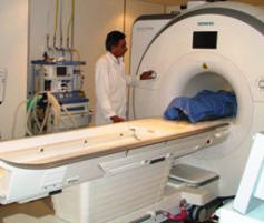
Skyra MRI with all clinical applications in the run since 28-Novemeber-2013.
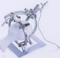
Inomed Riechert-Mundinger System, with three point
fixation is the most accurate system in the market. The microdrive and
its sensor gives feed back about the localization.
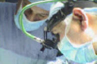
Inomed MER system
Leica HM500
The World's first and the only Headmounted Microscope.
Freedom combined with Outstanding Vision, but very bad video recording and
documentation.
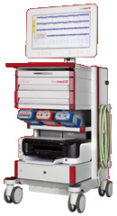
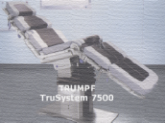
After long years TRUMPF TruSystem 7500 is running with in the neurosuite at
Shmaisani hospital starting from 23-March-2014
Fig-1: Bony deformity in AP view reconstructed using ORS Visual.
Fig-2: Bony deformity in left oblique view reconstructed using ORS
Visual.
Fig-3: Bony deformity in right oblique view reconstructed using ORS
Visual.
Fig-4: The Lynch Howarth incision.
Notice: Not all operative activities
can be recorded due to lack of time.
Notice: Head injuries and very urgent surgeries are also
escaped from the plan .