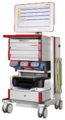
Inomed Stockert Neuro N50. A versatile
RF lesion generator and stimulator for
countless applications and many uses
Multigen RF lesion generator .
AMIRA MAHER AL-SHAREEF 28 YEARS
PERSISTENT CSF LEAK FROM THE CRANIOTOMY SITE.
Anamnesis
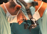
The patient was operated by me
10-May-2026
for multiple arachnoid cysts left cerebral
hemisphere and fenestration of the arachnoids
was done. The craniotomy site started to show
pocket of CSF under the wound, which with time
started to increase until the last 3 days 42 ml
was evacuated twice daily and the bone over the
craniotomy site came out. The patient is
deteriorating, and convulsions took place the
last day for what surgery was planned tomorrow.
After putting Foleys catheter and starting
Mannitol and Decadron, the CSF leak
stopped at the night before operation.
The craniotomy was revised
and the tense brain shifted the bone outside the
skull. The dura was tense and it was needed to
give the patient Mannitol and hyperventilation
with elevation of the head to obtain acceptable
feature to fit the bone in place with using the
PALACOS R+G bone cement to obtain water-tight
closure of the wound to avoid any CSF leak from
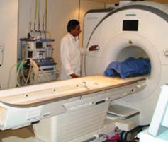
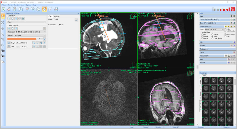
this area. Using intraoperative MRI with ceramic
ring of RM frame with U version of the fudicials
and using the IPs 6 software, the target and
entry points were obtained. According to target
point the distance was 94 mm from the surface of
bone. It was supposed to reach the pineal body.
Mini bore hole was created at the entry point
and the external drain was inserted. The
Medtronic external drain was used. The CSF came
out crystal clear. The operation in 2
sessions took about 8 hours, because the
fudicials were almost missing and it was to work
around to find the target point and entry points
without the complete package of IPs 6.
The patient start to recover
and sent to the ward. The CSF is crystal clean
and pulsating.
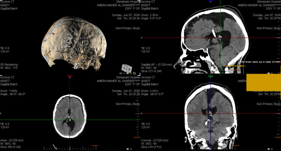
CT-scan was done 4 hours
after recover, to find that the tip of the drain
is in the right quadrigeminal cistern. The was
due to upward migration of the brain after
release of the pressure.
FOLLOW UP
The patient planned to
undergo permanent shunt using Medtronic
medium pressure after 2 days with retracting the
tip of the catheter 15 mm above.
Comments
The case is very complicated, because
there are no previous MRI data to compare with.
The VPS has small reservoir, and not
functioning for unknown period.
The family have controversial
information, that the surgeon cannot configure what is going
on, but the Jacksonian attack of the right upper limb, which
took place 2 years ago and the the drop right foot, can
denote that the seizure activity took place from the leg and
hand area of the sensomotor strip left hemisphere.
This case make it clear to not reach the
pineal body and to keep at least 15 mm above the target to
avoid shifting of the brain after decompression and upward
migration of the brain stem.
Do not leave loops outside the bur hole
with no proper fixation to prevent slippage of the tube
intracranially.
Skyra MRI with all clinical applications in the run since 28-Novemeber-2013.
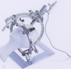
Inomed Riechert-Mundinger System, with three point
fixation is the most accurate system in the market. The microdrive and
its sensor gives feed back about the localization.
Inomed MER system
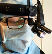
Leica HM500
The World's first and the only Head mounted Microscope.
Freedom combined with Outstanding Vision, but very bad video recording and
documentation.
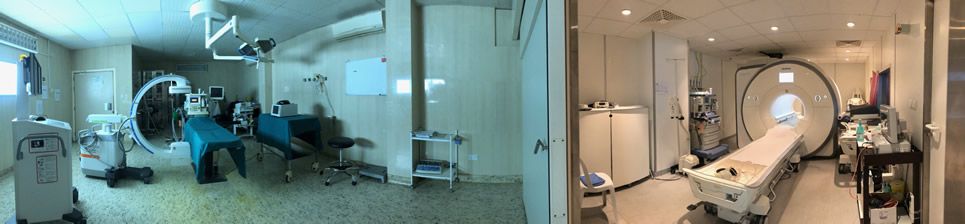
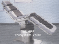
After long years TRUMPF TruSystem 7500 is running with in the neurosuite at
Shmaisani hospital starting from 23-March-2014
LooksCam II Xenosys in the run starting from 14-March-2021 with
SheerVision TTL x4 magnification.
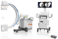
Cios-Spin flat panel in the run.
IPS 6 Inomed software showing the the entry and target points.
Notice the depth of the intracranial part is 96 mm.
The external drain after insertion for 96 mm by calculations by
Inomed software pierced near the pineal body in the right
quadrigeminal cistern for 30 mm.
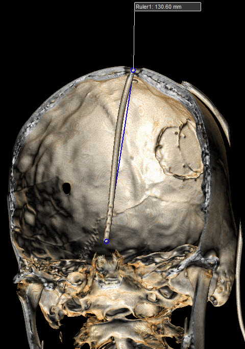
By proper measurement the depth of the intracranial part if the
device is 130 mm, which means the loop around the bur hole,
disappeared, which was the cause of deep penetration beyond the
target point.
Notice: Not all operative activities
can be recorded due to lack of time.
Notice: Head injuries and very urgent surgeries are also
escaped from the plan .