|
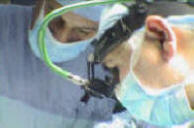
Functional Neurosurgery
functionalneuro.surgery
Functionalneurosurgery.net
IOM Sites
iomonitoring.org
operativemonitoring.com
Neurosurgical Sites
neurosurgery.art
neurosurgery.me
neurosurgery.mx
skullbase.surgery
Neurosurgical Encyclopedia
neurosurgicalencyclopedia.org
Neurooncological Sites
acousticschwannoma.com
craniopharyngiomas.com
ependymomas.com
gliomas.info
gliomas.uk
meningiomas.org
neurooncology.me
pinealomas.com
pituitaryadenomas.com
Neuroanatomical Sites
humanneuroanatomy.com
microneuroanatomy.com
Neuroanesthesia Sites
neuro-anesthessia.org
Neurobiological Sites
humanneurobiology.com
Neurohistopathological
neurorhistopathology.com
Neuro ICU Site
neuroicu.info
Neuroophthalmological
neuroophthalmology.org
Neurophysiological Sites
humanneurophysiology.com
Neuroradiological Sites
neuroradiology.today
NeuroSience Sites
neuro.science
Neurovascular Sites
vascularneurosurgery.com
Personal Sites
cns.clinic
Spine Surgery Sites
spine.surgery
spondylolisthesis.info
paraplegia.today
Stem Cell Therapy Site
neurostemcell.com

Inomed Stockert Neuro N50. A versatile
RF lesion generator and stimulator for
countless applications and many uses

Multigen RF lesion generator .
|
INTRACRANIAL
VASCULAR MALFORMATIONS
Historical Background
Intracranial arteriovenous malformations were studied and classified as
early as the mid-1800s (Luschka, 1854: Virchow. 1863). with the first
surgical exposure of an arteriovenous malformation by Giordano occurring
about three decades later in 1890. Fedor Krause attempted to surgically
eliminate an arteriovenous malformation by ligating its feeding arteries in
1908 but Olivecrona appear, to have been the first to actually completely
excise a cerebral arteriovenous malformation (AVM) in 1932 and later a
cerebellar AVM in 1938. Except at a few major centers. however, an
aggressive surgical approach to the larger examples of these lesions have
awaited the major technological advances of neurological surgery,
neuroradiology, and neuroanesthesia during the past several decades.
Embryology of Arteriovenous Malformations
Arteriovenous malformation of the brain are congenital lesions most likely
developing during the late somite stages of the fourth week of embryonic
life and almost certainly no later than the eighth week. The primary
pathologic lesion consists of one or more persisting direct connections
between the arterial inflow and venous outflow without an intervening
capillary bed.
Early in the third week of embryonic life, cells (angioblasts) begin to
differentiate from the mesoderm, forming small, syncytial islands. These
small clumps of syncytial cells develop tiny sprouts that extend to
interconnect the cell groups, forming a syncytial plexus. Intercellular
clefts appear within the syncytial masses. These clefts fuse to form the
primitive vascular lumen. The syncytial cells enveloping these clefts become
the endothelium of the new vessels. Proliferative growth of this endothelium
links the vascular lumina into a continuous irregular endothelial vascular
meshwork over the surface of the developing brain. Further extension of the
primitive network, present over the developing telencephalon of human
embryos at 4 weeks of age, occurs through endothelial sprouting.
Sabin has described a fascinating alternative process for the development of
the primitive vascular plexus. She observed the appearance of intracellular
vacuoles which coalesced to form the future vascular lumen, with the liquid
of the vacuole becoming the primitive plasma. According to this schema, the
first primitive vascular lumen is embryologically an intracellular
structure, with the syncytial cell, containing these interconnected vacuoles
forming the primitive vascular endothelium.
The primordial vascular plexus first differentiates into afferent, efferent,
and capillary components over the more rostral portion of the embryonic
brain. The more superficial portion of the plexus forms larger vascular
channels. eventually evolving into the arteries and veins, with the deeper
portion resolving into the capillary component more closely attached to the
brain surface. Beginning circulation to the brain appears around the end of
the fourth week of embryonic life. Arteriovenous malformations arise from
persistent direct connections between the future arterial and venous sides
of the primitive vascular plexus, with failure to develop an interposed
capillary network .
During the sixth and seventh weeks the third pair of aortic arches, together
with the dorsal aorta, transform into the primitive internal carotid
arteries, with the first and second arches undergoing early involution. The
vertebral arteries arise from a longitudinal linkage of the dorsal rami of
the intersegmental arteries of the neck during the fourth week. All the
original proximal intersegmental artery stalks except the most caudal one
atrophy, resulting in a longitudinal vessel taking origin along with the
subclavian from the sixth cervical intersegmental artery . The vertebral
artery establishes communication with the internal carotids through the
basilar artery, which arises independently through the consolidation of two
longitudinal vascular channels beneath the brain. This linkage is
established by the sixth week of fetal life. Between the sixth and eighth
week of fetal life, a compartmentalized brain, dural and extracranial
circulation has been established. By the eighth week of fetal life the major
venous sinus pattern of the adult has begun to emerge.
Pathologic Classification of Arteriovenous Malformations
The development of cerebral angiography catalyzed interest in the study of
intracranial vascular anomalies, providing the first major new insights into
the pathophysiology of these lesions. The first major classifications of
intracranial vascular malformations used extensively in the older European
literature, consisted of four overall categories: (1) angioma cavernosum,
(2) angioma racemosum, (3) angioreticuloma and (4) angioglioma. Angioma
racemosum included the subheadings of (a) telangiectasis, (b) Sturge-Weber
syndrome, (c) angioma racemosum arteriale, (d) angioma racemosum venosum.
and (e) arteriovenous aneurysm. The term "arteriovenous aneurysm"
corresponds to the current designation "arteriovenous malformation."
In 1966 McCormick proposed a more clinically oriented categorization into
five pathologic types: (1) telangiectasia, (2) varix, (3) cavernous angioma,
(4) arteriovenous malformation and (5) venous angioma. Telangiectasias are
capillary angiomas, usually small and solitary and most frequently occurring
in the pons and the roof of the fourth ventricle. They are only occasionally
associated with hemorrhage. A varix is usually quite small and is
occasionally invisible grossly, consisting of one or more dilated veins not
associated with an arteriovenous shunt. These small lesions, found in either
the parenchyma or the leptomeninges, may be associated with hemorrhage,
occasionally massive. Cavernous angiomas are dilated sinusoidal vascular
anomalies varying in size or diameter from 1 mm up to many centimeters and
are associated with hemorrhage as well as seizures. They occur most often in
the cerebrum but may occur in any part of the central nervous system. Brain
parenchyma is absent between the sinusoidal vascular spaces. Calcium
deposition and hyalinization of the vessel walls are common: spontaneous
thrombosis of either part or all of the lesion may occur. The blood in a
cavernous angioma is not arterialized. The term venous angioma defines a
malformation consisting entirely of veins not associated with an
arteriovenous shunt, though otherwise closely resembling an arteriovenous
malformation in gross appearance.
The term arteriovenous malformation, the primary topic here, refers to a
congenital maldevelopment of blood vessels, with preservation of one or more
primitive direct communications between arterial and venous channels. The
malformations are found throughout the central nervous system, occurring
most commonly in the cerebral hemispheres, with from 70 to 93 percent found
in the supratentorial structures in various reported series. Arteriovenous
malformations of the cerebral hemispheres most frequently involve the
distribution of the middle cerebral arterial tree, followed in declining
frequency by those of the anterior and then the posterior cerebral arteries.
Hemispheral arteriovenous malformations can be further subclassified into
those involving either one or a combination of the epicerebral, the
transcerebral, and the subependymal circulations.
The epicerebral circulation consists of short perforating branches arising
from the small pial arteries on the cortical surface and penetrating the
cortex more or less at right angles to the brain surface. They form a
distinct palisade of parallel short arteries of varying length, supplying
the superficial, middle, and deep layers of the cortex. These slender
cortical arteries show a grapnel-like pattern of branching, spreading
outward and back upward toward the cortical surface as they terminate in a
capillary bed. The longer transcerebral arteries (averaging 2 to 3 cm in
length), traverse the cortex to feed an elongated capillary mesh or plexus
paralleling the transcerebral arteries in the white matter. The
transcerebral arteries terminate in the periventricular plexus.
Paralleling the arterial pattern, the venous drainage of the epicerebral
circulation courses back outward to the veins on the pial surface. The
venous drainage of the transcerebral arterial circulation is predominantly
inward toward the subependymal venous plexus of the lateral ventricles,
though anastomotic connections with and associated flow to the epicerebral
veins are also present.
Malformations involving only the transcerebral arteries are not visible on
the cortical surface, although it is common to see arterialized venous
channels on the pial surface of the cortex as a result of the anastomotic
connections between the transcerebral and epicerebral venous drainages.
Pathology
The gross appearance of an arteriovenous malformation is that of a tangled
mass of dilated tortuous vessels. Small areas of hemosiderin staining and
thickened, milky appearing pia-arachnoid are common in the immediate
vicinity of the lesion in older patients. If the transcerebral circulation
is involved in the malformation, the lesion presents a characteristic
wedge-shaped appearance with the apex of the wedge at the ependymal surface
of the lateral ventricle and the base of the wedge parallel to the overlying
cerebral convexity. There is a rare but surgically very favorable group of
arteriovenous malformations limited entirely to the pial surface of the
brain stem.
Arteries emptying into the malformation become passively enlarged with time
due to the high flow volume resulting from the abnormally low peripheral
resistance of the A-V shunt. The venous system draining the shunt similarly
undergoes progressive enlargement with increasing tortuosity as a result of
the high flow volume and sustained increased venous pressure produced by the
A-V shunt. Atrophic changes of the cortex and subcortical white matter in
the immediate vicinity of the malformation are also common findings in older
patients. Secondary changes with time have been found in the arterial walls
of the feeding arteries in the immediate vicinity of the malformation, with
collagenous replacement of the normal smooth muscle component of the media.
Saccular aneurysms are an associated finding in between 10 and 15 percent of
patients with arteriovenous malformations. Between 60 and 95 percent of
these aneurysms occur on arteries hemodynamically related to the
arteriovenous malformation.
The external carotid artery may make a significant flow contribution to a
cerebral arteriovenous malformation and occasionally may be the sole source
of arterial inflow to the lesion.
Incidence: Age and Sex Distribution
The cooperative study on intracranial aneurysms and arteriovenous
malformations suggested that the frequency of intracranial arteriovenous
malformations is about one-seventh that of saccular aneurysms. This would
indicate that about 0.14 percent of the population harbor one of these
lesions in a given year. The majority of lesions become symptomatic by the
age of 40 and in most large series show no predilection for either sex.
Although occasional reports of familial incidence are found in the
literature, the larger series show no familial or genetic predisposition.
Clinical Features
In adult life the first symptom of an arteriovenous malformation is usually
either a hemorrhage or a seizure, These two types of presentation occur with
about equal frequency, The average age of onset for epilepsy as the initial
symptom is about age 25, with age 30 the corresponding figure for
hemorrhage. Patients with large arteriovenous malformations are more than
twice as likely to have seizures in contrast to hemorrhage as their initial
symptom, whereas the reverse is found for small lesions.
The reported incidence of headache from an arteriovenous malformation as an
early symptom before the onset of either seizures or a hemorrhage range,
from 5 to 35 percent. A pseudotumor syndrome secondary to elevated venous
sinus pressure from large A-V shunts, particularly if the shunts are near
the torcular and transverse sinus, and hydrocephalus as a sequelae to
previously undiagnosed small subarachnoid hemorrhages are less common as a
presenting feature. Arteriovenous malformations may occasionally mimic a
demyelinating disease or brain tumor, particularly, when located in the
brain stem or deep basal ganglia. Intellectual deterioration tends to occur
with large AVMs in the older age groups. This deterioration appears to be at
least partially related to a cerebral steal phenornenon.
In children. hemorrhage is seven times more likely than a seizure to be the
initial presenting event. An additional common presentation of an
arteriovenous malformation in the neonatal period is high-output left
ventricular cardiac failure. Detailed hemodynamic studies have shown that
right heart failure may evolve as an additional complicating factor
secondary to right side overload from the left to right shunt.
The clinical course of an arteriovenous malformation, apart from hemorrhage,
is usually one of slowly progressing symptomatology referable to the site of
the lesion. The mortality rate from hemorrhage in the cooperative study was
10 percent from the initial bleeding episode. 13 percent from a second
episode, and 20 percent from a third episode. The risk of recurrent
hemorrhage after an initial bleeding episode is between 3.5 and 4.0 percent
per year. The risk of hemorrhage in a patient presenting with cerebral
seizures but with no known previous hemorrhage has been variously reported
as between 1 and 2.3 percent per year. Forster et al. found. in a 15-year
average follow-up of 35 patients presenting with epilepsy alone, a 17
percent mortality and 20 percent severe disability secondary to hemorrhage.
They further noted that if the patient had had one hemorrhage, there was a
25 percent risk of rebleeding over the next 4 years. If there had been two
previous hemorrhages, the risk for further rebleeding was 25 percent within
the year following the most recent hemorrhage. A review of 137 patients
treated conservatively with a follow-up period ranging from a minimum of 10
years to a maximum of 25 years found that only 20 percent of the 137 were
alive and well at the end of the study. Thirty-seven patients either had
died or were severely incapacitated by the arteriovenous malformation.
Vascular malformations presenting during pregnancy are more likely to
rehemorrhage than those in the nonpregnant patients with the frequency of
rebleeding approaching that of saccular aneurysms. The posthemorrhage
mortality and morbidity figures, however, remain significantly lower than
those for saccular aneurysms and comparable with those for the nonpregnant
individual. Surprisingly, the timing of rebleeding does not appear to peak
or parallel the cardiovascular changes in pregnancy. The peak incidence of
hemorrhage from AVMs occurs between the fifteenth and twentieth week of
pregnancy as compared with the peak incidence of aneurysm rebleeding between
the thirteenth and fourteenth week of gestation. Only 2 of 77 AVM
hemorrhages during pregnancy in this series occurred during labor. Elective
cesarean section at 38 weeks gestation was thought to carry the smallest
combined risk to mother and child.
Occasional spontaneous disappearance of intracranial arteriovenous
malformations has been reported, but this remains a very rare occurrence.
Radiology
Cerebral angiography continues to be the definitive study for the assessment
of intracranial vascular malformations. Careful bilateral carotid as well as
vertebral angiography often demonstrates unexpected crossover or collateral
filling of AVMs and is essential for adequate planning of therapy and
assessment of risks to the patient. Computed tomography (CT) scanning or
magnetic resonance imaging (MRI) have become common screening techniques for
the diagnosis of vascular malformations. Angiographically occult AVMs have
been found using both imaging techniques. Intracerebral hemorrhage enhancing
on CT scan, even when arteriography fails to demonstrate a vascular anomaly,
should raise the suspicion of the presence of a small AVM. Neither CT nor
MRI reveals the anatomic detail necessary for surgical planning. They also
do not reliably disclose the presence of associated vascular anomalies such
as saccular aneurysms.
In a group of patients with AVMs studied with unenhanced, enhanced, and 1-h
postcontrast CT scans, the precontrast scan was abnormal in 81 percent of
patients. 2% of patients showed a venous angioma on the immediate
postcontrast scan, which was not apparent on either the precontrast or the
1-h delayed scan. The 1-h delayed scan revealed one angiographically occult,
thrombosed AVM not seen on the precontrast or immediate postcontrast scan.
The 1-h delayed scan also showed additional pathologic changes in areas
adjacent to the lesions shown on the precontrast and immediate postcontrast
scans. Delayed high-contrast CT scanning was judged to show no advantage as
the routine screening procedure and, if done as a sole procedure, might miss
at least some venous angiomas.
The "flow void" seen on MRI of AVMs has become a useful, though not
completely accurate, technique for assessing the degree of occlusion of AVMs
after focused stereotactic radiation therapy.
Indications for Operation
The role of surgery in the clinical management of a given patient is based
on a composite of the probable natural history of the patients future
clinical course, the risk of surgical management with particular reference
to the patient's required occupational or daily activities, and finally, the
patient's age. Patients in the older age group who have seizures but who are
otherwise neurologically intact and without a previous history of hemorrhage
have comparatively a smaller cumulative risk of major morbidity and
mortality with continued conservative management. An important factor in
long-term planning for the younger patient is the problem that seizure foci
secondary to AVMs tend to become progressively more resistant to medical
management with time. Although most current surgical series show some
reduction in seizure tendency after malformation excision, extirpation of
the malformation more importantly may block the further development of
medically intractable seizure activity. In the younger patient, as is
discussed in more detail below, the risk of mortality or major morbidity
with surgery using current techniques is competitive with the 10-year
prognosis for lesions that have not bled, and is better than the 5-year
prognosis for malformations with a previous history of at least one
hemorrhage. Malformations in areas of eloquent function are being found
increasingly amenable to a surgical approach, with mortality or major
morbidity risks of 10 percent or less. Deep lesions involving the internal
capsule, thalamus, midbrain and lower brain stem are still usually found to
be inoperable in terms of acceptable risks to neurological function.
Role of Embolization in AVM Management
Embolization of larger AVMs has become an important therapeutic adjunct to
their surgical management. To date, the large majority of these lesions
cannot be totally occluded by embolization techniques. Embolization does,
however, permit a staged preoperative reduction in size of the arteriovenous
shunt, producing significant circulatory readjustment and reducing the
degree of hydraulic shock resulting from the final occlusion of the fistula
at the time of surgical resection of the lesion. Embolization, when
practical, has largely replaced staged surgical occlusion of the feeding
arteries to achieve this effect.
Embolic agents are classified as either absorbable or nonabsorbable and as
either solid or fluid. Solid embolic agents have been injected into the
internal carotid or vertebral artery feeding the malformation, relying on
the high-volume axial flow characteristics of the circulation to the AVM to
carry the solid particles into its nidus. This technique is not satisfactory
if the pellets, such as nonabsorbable barium-impregnated silicone spheres,
have to leave the parent artery at a sharp angle to enter a branching
vessel, such as would be required for a pellet entering the anterior
cerebral artery from the internal carotid artery.
Gelfoam, cut into 1 x 2 mm strips, impregnated with tantalum powder and
soaked in angiographic contrast material has been a common absorbable solid
embolic agent. Although this material is relatively easy to handle. it has
been more unpredictable in producing occlusion on the arterial side of the
shunt and has no major advantages over silicone spheres.
Fluid embolic agents that have been employed have been nonabsorbable and of
either the bucrylate or silicone types. Isobutyl-2cyanoacrylate (ICBA) is a
prototypic material of the bucrylate group. It is a rapidly polymerizing,
low-viscosity tissue adhesive which is made radiopaque by adding tantalum
powder. ICBA polymerizes rapidly on contact with ionic solutions such as
blood or normal saline, while a 5% glucose solution will block
polymerization. Considerable skill and experience are required in the use of
this material. The speed of polymerization and rate of injection must be
finely calculated to ensure that polymerization occurs on the arterial side
of the malformation. Distal migration of this fluid into the major sinuses
has occurred. If the arterial inflow is not arrested by polymerization on
the arterial side of the shunt, sudden swelling and rupture of the
malformation with major hemorrhage may occur. Bucrylate produces a foreign
body giant cell reaction with chronic inflammatory changes not only in the
vessel wall but also to a lesser degree in the adjacent brain parenchyma.
The long-term effects of this material are not yet fully known, Occasional
malformations have been completely occluded with bucrylate, although the
success rate for total occlusion has not been high, There are several
additional technical problems in using this material for occlusion of
malformations in areas of eloquent function, Arterial branches to normally
functioning eloquent cortex often depart from the parent artery distal to
the first arterial branches going to the malformation. Total occlusion of
the malformation would, of necessity, require sacrificing these normal
branches, with potentially serious neurological sequelae. Additionally, the
hardened, noncornpressible prongs of bucrylate within an incompletely
occluded malformation may significantly increase the difficulty of
subsequent safe separation and surgical removal of the malformation from
areas of critical function.
Silicone fluid mixtures have occasionally been used instead of bucrylate.
The mixture consists of a silastic elastomer containing a filler necessary
for vulcanization. and a medical-grade silicone fluid that acts as the
diluent to the more viscous Silastic elastomer. These two silicones are
mixed to the desired viscosity and then tantalum powder is added to permit
radiographic visualization. A catalyst to produce vulcanization is required.
The Silastic is injected just before vulcanization occurs. It has no
adhesive properties, so that a complete filling or cast of the vascular
lumen is required.
After embolization with attendant reduction in the sump effect of the AVM,
some patients have been noted to show improvement in intellectual
performance, suggesting the correction of some degree of symptomatic
cerebral steal. Wolpert et al. found, however, that embolization had no
long-term effect on the progression of neurological symptoms or signs and no
effect on seizure frequency. Incomplete occlusion of the malformation by
embolization has not reduced or modified the natural history of the lesion
with respect to hemorrhage.
In 1971, Serbinenko reported the use of detachable flowdirected balloons on
the tips of catheters threaded into the proximal vessels to the
malformation. This technique has been a key factor in permitting selective
catheterization of these vessels for the injection of embolic agents but has
not been a satisfactory therapeutic occlusive maneuver in and of itself.
Operative Management
Preoperatively, the patient is placed on an anti epileptic to minimize the
risk of seizures during the early postoperative period of cerebral
vasocongestion and cerebral swelling, even if the patient has no previous
history of cerebral seizures. Serum antiepileptic levels are checked
immediately before surgery to ensure that adequate antiepileptic levels are
present. Dexamethasone is started 36 to 48 h preoperatively to help
stabilize capillary membrane permeability during the early postoperative
interval of hydraulic shock and local tissue reaction to surgical
manipulation.
If the malformation lies in or immediately adjacent to the expected location
of the motor cortex or major speech centers, the surgical procedure may be
carried out under local anesthesia with cortical mapping to ensure accurate
localization of the areas of eloquent function and to permit the testing of
these functions serially throughout the removal of the malformation. In this
latter situation, temporary clips are placed on the arterial feeders
immediately proximal to the malformation, followed by function testing. The
temporary clips are then replaced with permanent ones if no functional
impairment has ensued.
Surgical resection should always be performed under magnification with
appropriate microsurgical instrumentation. The dissection plane follows
along the immediate margin of the malformation in the thin, gliotic
nonfunctional zone between the malformation and the adjacent cortex and
white matter. Particular care must be taken in occluding the small,
thin-walled endothelial tubules composing the transcerebral venous drainage.
These vessels are extremely fragile and, if torn, back-bleed profusely due
to the increased venous pressure in the subependymal venous plexus from the
A-V shunt. It is essential to avoid pursuing these vessels if unacceptable
neurological deficit is to be avoided. Temporary placement of small fluffy
cotton pledgets, accompanied by surgeon patience and by moving on to another
area of the removal, will normally secure hemostasis of these individual
venous bleeding points. Careful positioning of the head so that the major
intracranial venous drainage is above heart level is a major factor in
reducing venous congestion and attendant blood loss.
Selective identification and occlusion of the arterial inflow to the lesion
with protection of the venous drainage as long as possible is important,
although Malis advocates using one of the draining veins as a 'handle" and a
guide to resection when several major draining wins are present. Major
reduction in venous outflow before interruption of the arterial inflow must
be avoided if malformation rupture with massive bleeding is to be avoided.
High-contrast visual dye can be injected intra-arterially to aid in the
identification of the feeding arteries to the malformation if the vascular
tangle of the malformation makes selective identification of the arterial
inflow otherwise difficult. Significant fragility of the lesion persists
down to the very end of the resection, making it essential that neither
fatigue nor impatience results in a rush or hurry to complete the final
stages of the removal.
A grid technique of localization of cortical function for a malformation
lying in or adjacent to the central areas has been proposed by Kune. This
technique presupposes a consistent pattern of cortical function with
reference to standard anatomic landmarks. Experience with cortical mapping
unfortunately has revealed significant deviation in location from the more
common patterns of cortical function around the margins of arteriovenous
malformations, especially with respect to speech localization. Modern
techniques of anesthesiology have made a major contribution to increasing
the safety of the surgical approach to,. and manipulation of these lesions.
Moderate hypotension during critical periods of surgical resection is well
tolerated, even under local anesthesia and does not interfere with patient
alertness and function testing. The general anesthesia technique of jet
ventilation can also essentially eliminate brain movement secondary to
respiration.
Preliminary experience with surgical lasers used on intracranial vascular
lesions has appeared in the literature. At present, the lasers seem to have
limited application to the surgery of AVMs. This is particularly true of the
CO2 laser, which has relatively poor vessel coagulation ability because of
its extremely shallow depth of penetration. The CO2 laser tends to punch
holes in the walls of larger vessels. The neodymium:YAG laser is more
efficient in achieving hemostasis due to its greater depth or penetration.
However, this latter laser type also is not effective in providing adequate
hemostasis in dealing with the very thin-walled endothelial tubes of the
engorged transcerebral circulation The neodymium:YAG laser appear, to
provide adequate vascular occlusion when contractile elements are a
significant component of the vessel walls being treated with the laser.
Gentle handling of the arteries proximal to the lesions is essential,
particularly in the posterior fossa where proximal propagation of clot from
the point of arterial occlusion can result In a disastrous outcome for an
otherwise technically satisfactory surgical excision. After completion of
the resection, the patient's blood pressure should be brought to normal
levels and the operative field observed carefully to ensure that hemostasis
is complete. Feeding arteries of 1 mm or larger must be securely clipped, if
delayed postoperative hemorrhage is to be consistently avoided. Bipolar
coagulation alone for these larger vessels is not adequate.
Postoperatively, the patient is nursed with the head of the bed elevated 30
to 40 degrees to maintain optimal venous outflow. It is helpful to maintain
the systolic blood pressure between 90 to 110 mmHg. using a trimethaphan
camsylate drip, to minimize the effects of hydraulic shock and attendant
hyperperfusion around the margins of the resection during the first 24 h
postoperatively. Crystalloids are restricted in order to produce a mild
dehydration, with the goal of a serum osmolarity between 295 and 305. Blood
volume is maintained with colloid administration. Dexamethasone is continued
postoperatively for 8 to 10 days and is then rapidly tapered. Postoperative
angiography is essential to confirm that complete removal of the
malformation has been achieved.
Results
The type of patient screening before surgical referral as well as the
aggressiveness of the consulting neurological and neurosurgical units are
obvious factors in reported results. In larger series in which over 60
percent of all patient, referred underwent surgical extirpation of the
lesions, a mortality rate ranging from 7 to 14 percent is found. The
widespread use of the surgical microscope and the staged preoperative
embolization of the lesions are major factor, in the improving mortality and
morbidity statistics. Surgical mortality rates now appear to compare
favorably with the long-term mortality rates of these lesions managed
conservatively in the younger patient. More information regarding the
quality of postoperative survival, as compared to the quality of life with
conservative management. is needed, In the few instances where this
information is beginning to appear, preliminary indications are that the
long-term quality of life is more favorable when surgical extirpation of the
lesion has been carried out.
|
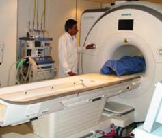 Skyra MRI with all clinical applications in the run since 28-Novemeber-2013.
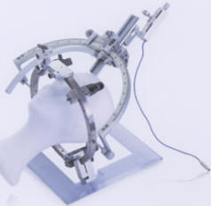
Inomed Riechert-Mundinger System, with three point
fixation is the most accurate system in the market. The microdrive and
its sensor gives feed

MER Inomed system.

Inomed ISIS IOM
highline with 32 channel and Neuroexplorer version 5 is functioning for
several months, starting from 01-August-2007. For more detailed information
about this functional neuronavigation machine with its early alarming signals,
please refer to inomed.com
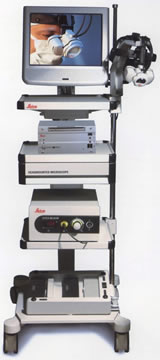
Leica HM500
The World's first and the only Head mounted Microscope.
Freedom combined with Outstanding Vision, but very bad video recording and
documentation.

After long years TRUMPF TruSystem 7500 is running with in the neurosuite at
Shmaisani hospital starting from 23-March-2014
|


