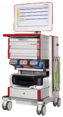
Inomed Stockert Neuro N50. A versatile
RF lesion generator and stimulator for
countless applications and many uses
Multigen RF lesion generator .
16-OCTOBER-2021 BASEM MUHAMED MAREEKH 31 YEARS
CONGENITAL BILATERAL ISTHMOLYSIS L5-S1.
Anamnesis
The patient came to the clinic 18-May-2019
complaining of chronic LBP since he was 14 years
age with intermittent course. 5 years later he
got left sciatica. The last 8 months got
bilateral sciatica more the left. MRI lumbar
spine performed 15-May-2019 showed bilateral
L5-S1 isthmolysis with fluid in the L5-S1
joints.
On examination, the patient in agonizing pain,
limping with exaggerated scoliotic stance. SLRS
85 degrees right side with pain and 85 degrees
left side with pain. There is no sensory nor
motor deficit.
The patient was sent for investigations and
dynamic studies showed I degree
spondylolisthesis and CT-scan constructed with
ORSVisual showed bilateral L5-S1 isthmolysis.
The patient then came 26-August-2021 with
dramatic deterioration the last 20 days with
exacerbation of the left sciatica and weak
dorsiflexion left foot 4/5 and SLRS was 80
degrees both sides with pain. The investigations
repeated and the same data were achieved.
Skeletonization of L5, lower
half of L4 and upper sacrum the lateral
processes of L5 were seen. Using
ERISMA-LP EVOLUTION system, transpedicular
screws, polyaxial to upper sacrum and monoaxial
to L5 body were inserted 6x40 dimensions. Using
MultiGen the right screws and lower left
responded brisk at 4.0 Volts. Foraminotomy both
S1 roots showed that the right S1 screw is in
bone but 2 mm near the medial wall, for what the
screw was reinserted 5 mm lateral to the first.
Motor stimulation still showing response but to
less degree. The root is away from the screw
shaft. Bipolar stimulation of the right S1 root
was achieved with 0.3 Volts. It became clear
that the root is abnormally responding to any
stimulation and the screw is far enough. The
left lower screw also repositioned 5 mm lateral
and no response to 4 Volts. The right L5 screw
was also repositioned lateral and no monopolar
stimulation even with 4 Volts. Using
bended rods fusion of the area was achieved and
the bony material of the removed flail lamina
was used lateral to the rods. The lamina of of L5
is flail, it was totally removed. Using
MultiGen, bipolar motor stimulation of the right
S1 root was achieved with 0.3 Volts. The left
responded to 0.7 Volts. A
bipolar pulsed mode RF with 42 Celsius, 240 sec,
2 Hz and 20 msec duration to both S1 roots
was achieved using 4 bended catheters 10 mm
exposed length. Further bipolar motor
stimulation of the right
L5 root was achieved with 0.3 Volt and the left
with 0.6 Volt. The patient was put in Reverse
Trendelenburg position with Valsalva maneuver
and hyperventilation. No CSF leak. A fat tissue
with pedicle was used to cover the dura to minimize the postoperative scar
formation and prevent postoperative CSF leak.
Routine closure of the wound. Smooth
postoperative recovery. The power of the left
foot improved.
He was sent to the
ward.
MultiGen
FOLLOW UP
Too early now.
Comments
This is the 227th case using the BPRF mode
with MultiGen. This procedure regained routine acceptance.
It became a usual part of the spine and peripheral nerves
surgery. Click here for
reference.
It still unclear to evaluate the
differences of pre and post application motor responses. The
only sure thing that it tells that the electrodes did not
migrate during the procedure and the nerve is functioning
properly. Here the threshold of motor
stimulation of the affected nerve showed dramatic
improvement after BPRF.
With accumulation of data, it became
clear that the irritated nerve with aberrant currents
running in the C fibers up, not only causing no change or elevation of
the required voltage to achieve motor response, but they could cause the preoperative
weakness. Ablation of such currents results in facilitation
of the motor response and improvement of function with
disappearance of pain.
It is unclear why the roots have several
motor response with different patients, despite the fact
that the neurological status is the same and the anesthesia
protocol also the same.
It could be that the nerve is recovering
minute by minute after decompression and this can explain
why the motor conductivity is improving after the BPRF
application, which require 4 minute session in most cases.
After the 172d case, the elevation of
motor stimulation above 5 V was abandoned to avoid delayed
dural tear with subsequent CSF leak, which take place at the
contact at the lower electrode shaft with the dura below the
level of the axilla.
This case is a demonstration that in rare
cases when the root is responding to very small currents, it
could mislead the surgeon that the screws are violating the
medial wall of entry, despite the fact that the screws are
in acceptable position and forces the surgeon to reposition
the screws more far as in this case.
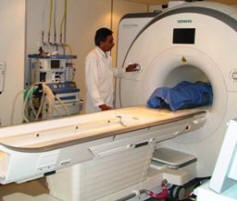
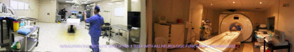
Skyra MRI with all clinical applications in the run since 28-Novemeber-2013.
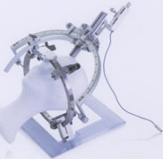
Inomed Riechert-Mundinger System, with three point
fixation is the most accurate system in the market. The microdrive and
its sensor gives feed back about the localization.
Inomed MER system
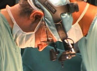
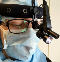
Leica HM500
The World's first and the only Head mounted Microscope.
Freedom combined with Outstanding Vision, but very bad video recording and
documentation.
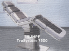
After long years TRUMPF TruSystem 7500 is running with in the neurosuite at
Shmaisani hospital starting from 23-March-2014
LooksCam II Xenosys in the run starting from 14-March-2021 with
SheerVision TTL x4 magnification.
Notice: Not all operative activities
can be recorded due to lack of time.
Notice: Head injuries and very urgent surgeries are also
escaped from the plan .