Munir Elias 20-12-2013

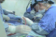
Most of the site will reflect the ongoing surgical activity of Prof. Munir Elias MD., PhD. with brief slides and weekly activity. For reference to the academic and theoretical part, you are welcome to visit  neurosurgery.tv

Functional Neurosurgery
functionalneuro.surgery
Functionalneurosurgery.net

IOM Sites
iomonitoring.org
operativemonitoring.com

Neurosurgical Sites
neurosurgery.art
neurosurgery.me
neurosurgery.mx
skullbase.surgery

Neurosurgical Encyclopedia
neurosurgicalencyclopedia.org

Neurooncological Sites
acousticschwannoma.com
craniopharyngiomas.com
ependymomas.com
gliomas.info
meningiomas.org
neurooncology.me
pinealomas.com
pituitaryadenomas.com 

Neuroanatomical Sites
humanneuroanatomy.com 
microneuroanatomy.com

Neuroanesthesia Sites
neuro-anesthessia.org

Neurobiological Sites
humanneurobiology.com

Neurohistopathological
neurorhistopathology.com

Neuro ICU Site
neuroicu.info

Neuroophthalmological
neuroophthalmology.org

Neurophysiological Sites
humanneurophysiology.com

Neuroradiological Sites
neuroradiology.today

NeuroSience Sites
neuro.science

Neurovascular Sites
vascularneurosurgery.com

Personal Sites
cns.clinic

Spine Surgery Sites
spine.surgery
spondylolisthesis.info
paraplegia.today

Stem Cell Therapy Site
neurostemcell.com


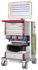
Inomed Stockert Neuro N50. A versatile
RF lesion generator and stimulator for
countless applications and many uses


Multigen RF lesion generator .

18-SEPTEMBER-2014  HIBA HAMED THAMER  30 YEARS  POSTERIOR THIRD OF THE III VENTRICLE TUMOR WITH SECONDARY HYDROCEPHALUS.


Anamnesis

bullet

The patient came to clinic 13-September-2014 complaining of headache for 2 months, bifronto-occipital localization without asymmetry associated with blurred vision and attacks of blindness, vomiting and episodes of nausea with attacks of de ja vu with fainting attacks. The condition is deteriorating. MRI of the brain done 07-September-2014 showing upper stem mass of bad quality, mostly posterior third of the III ventricle.

bullet

On examination; the patient is swaying in Romberg position. There is left hemihypalgesia with weak four limbs more the left side. The deep reflexes are more brisk in the left side. SLRS was 80 degrees in the left because of weakness.  The left quadriceps is -3/5 and the right is 3/5. The mother noting that the patient is hallucinating some times.

bullet

MRI of the brain with contrast and MRA, MRV of the brain with spectroscopy and DTI were performed the same day, showing the tumor inside the posterior third of the III ventricle, pushing upward the deep cerebral veins. The tumor is multi in consistency, but not invading the surrounding structures and pushing the mesencephalon down and the basal ganglii lateral.

bullet

Midline incision over the parietal and occipital area with craniotomy to include both sides of the postero-parietal and antero-occipital area. The dura was opened parallel to the sagittal sinus from the right and reflected to left.  Interhemispheric approach without scarifying any running vein. Transcallosal approach was achieved and the deep cerebral veins identified. The tumor was highly vascular and multiconsistency and was sent to frozen section which gave the result of highly malignant astrocytoma. The tumor was attacked until the bed of the mesencephalon was seen and the right lateral ventricle was under vision. It was felt that total resection was achieved. The patient was sent for MRI, which confirmed the presence of almost 2/3 of the tumor still persisting in the left side. The patient was brought back and the left part of the tumor was removed. Both lateral ventricles and the third ventricle and the preserved deep cerebral veins were seen. Inspection of the tumor cavity was inspected. All the surrounding wall are normal brain tissue. An external drain was left to the left lateral ventricle and routine closure of the wound was achieved.

bullet

Smooth postoperative recovery. The patient was extubated immediately after surgery and she is moving all limbs with good communication, but with episodes name disorientation.

Follow Up

bullet

The patient was doing well. When we started to taper the Decadron, she got mutism and with difficulty got verbal response, for what we elevated the Decadron to 8 mg three times a day, after what some improvement took place.

bullet

The final histologic result was anaplastic ganglioglioma.


Comments  

bullet

The patient without intraoperative MRI could be left with 2/3 of the tumor left behind. Do not ever trust your sense. Always check when you are in doubt, even when you are confident.

bullet

The patient will not need VPS since there is communication through the callosotomy site.

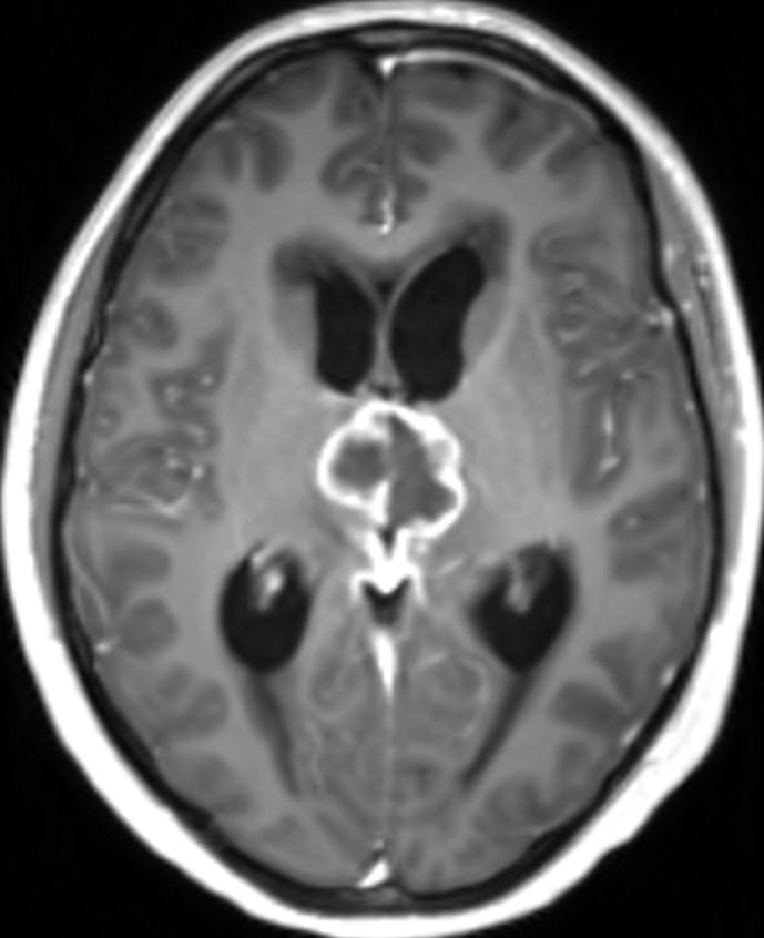
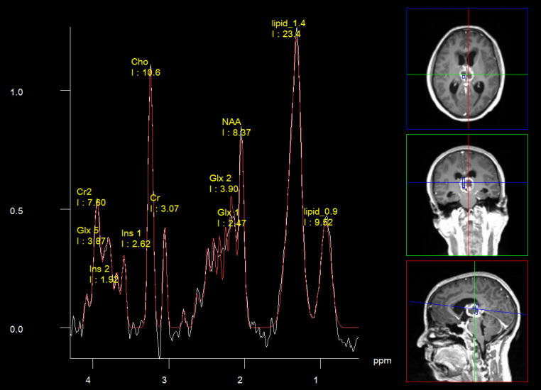
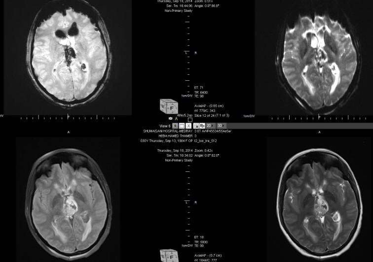
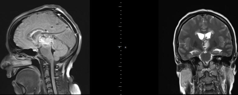
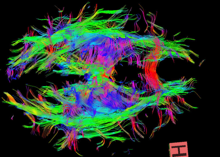
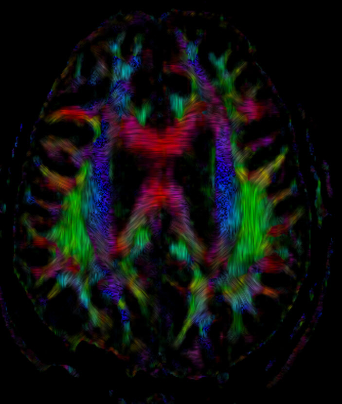
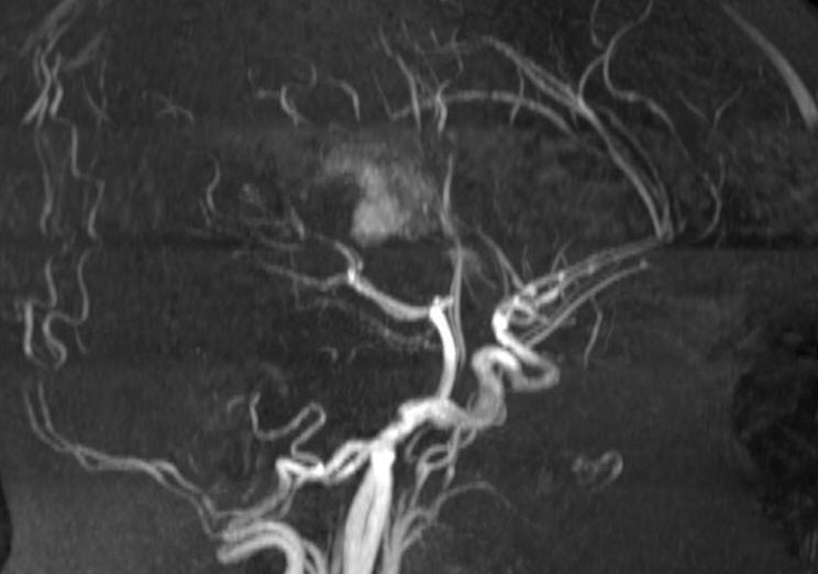
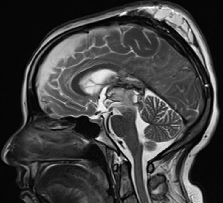
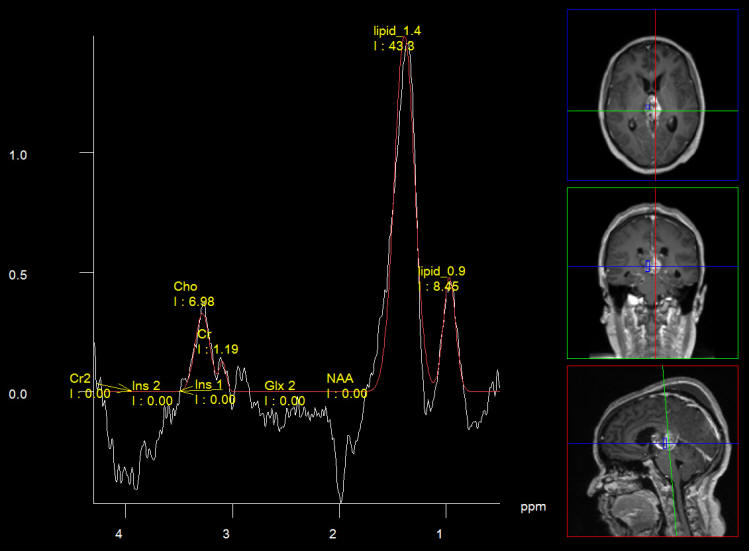
Skyra MRI with all clinical applications in the run since 28-Novemeber-2013.

Leica HM500

Leica HM500
The World's first and the only Headmounted Microscope.
Freedom combined with Outstanding Vision, but very bad video recording and documentation.

TRUMPF TruSystem 7500

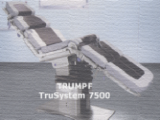
After long years TRUMPF TruSystem 7500 is running with in the neurosuite at Shmaisani hospital starting from 23-March-2014


Inomed MER system


Axial T1 with contrast.


Frontal T1 with contrast


Sagittal T1 with contrast.


Choline distribution showing a small nidus of possible malignant character.

 
NAA distribution.


Short Echo spectroscopy in favor of pinealoblastoma.


DTI showing no fibers inside the tumor mass.


Normal anatomy of the pineal gland


Same anatomy with reflected callosum.


The next intraoperative MRI showing total resection of the mass and floating venous structures with deformed choroidal veins.


Sagittal and coronal vies showing the external drain  and  a clot over the mesencephalon.


Postsurgical callosotomy due to transcallosal approach. Fibertraking done 27-September-2014


DTI showing the transcallosal approach in the posterior third. Done 27-September-2014.


Choline elevation confirming still persisting active sites intermingled with the hematoma in the right side.


Choline/NAA ratio confirming the presence of active lesion at the right side.


The hematoma in the bed of resected tumor.


Strict midline sagittal section showing the transcallosal approach and abundant of venous structures and the hematoma.


Spectroscopy short TE showing low choline ratio? with high lipids 1.3 and 0.9.

Back Up!

Notice: Not all operative activities can be recorded due to lack of time.
Notice: Head injuries and very urgent surgeries are also escaped from the plan .

WELCOME TO AL-SHMAISANI HOSPITAL


View Larger Map
 

© [2014] [CNS CLINIC - NEUROSURGERY - JORDAN]. All rights reserved