Most of the site will reflect the ongoing surgical activity of Prof. Munir Elias MD., PhD. with brief slides and weekly activity. For reference to the academic and theoretical part, you are welcome to visit
neurosurgery.tv
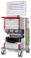
Inomed Stockert Neuro N50. A versatile
RF lesion generator and stimulator for
countless applications and many uses
Multigen RF lesion generator .
15-SEPTEMBER-2015 HUDA ABDEL-JALEEL AL-MBAYED 19
YEARS GLIOMA OF THE LEFT TEMPORAL LOBE.
Anamnesis
The patient came to the clinic 12-July-2015
complaining of epi attacks for 4 years and was
in Tegretol 400 CR twice a week. MRI of the
brain done 01-June-2014 showing cystic lesion
anterior to the left inferior horn.
On examination: The patient is alert with slight
right hemiparesis,
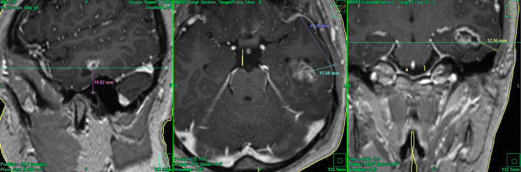
The patient was sent for new investigations and
MRI of the brain performed 22-July-2015 showing
the mass considerably enlarged in diameter
28.3x18.4 mm anterior to the left inferior horn.
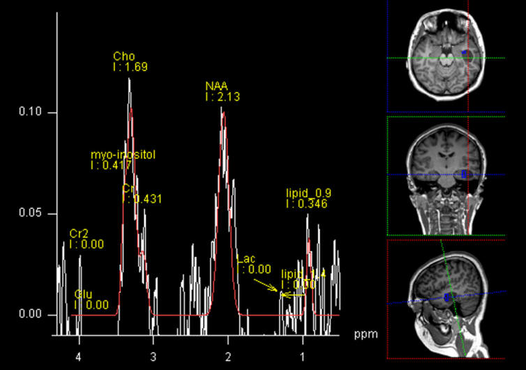
Spectroscopy showed high choline levels with
glioma nature of the mass.
Lazy S-shape incision to the
left temporal region. A bony flap was created
over the projection of the tumor. The dura was
opened in t-shaped fashion. Transcortical
approach to the tumor. The tumor was violate
friable, and multiple consistency. The tumor was
resected and the inferior horn was exposed as
landmark confirming the radical resection of the
mass, which was sent for histologic
investigations. Hemostasis and before closing
the wound, MRI was done, which confirmed the
radical resection of the mass but showing a
separate seeder in the left border of the
anterior commeasure abutting the posterior
border of the left MCA, reaching the left
Internal capsule. It was decided not to violate
them, because they are deeply seated and the
mortality and permanent postoperative plegia is
at best outcome. Routine closure of the
wound.
Smooth postoperative recovery. The patient
showed dense right sided plegia which was
improving over hours after surgery. She was
sent to the ward.
Follow Up
The patient showed mutism after surgery with
denial to move the right side of the body. She
could move with painful stimulation and produce
sounds. She could write and understand the
verbal commands. Largatil 25 mg was started
20-September-2015.
The final histologic result was astrocytoma
grade II.
Lesson
The data from which the plan was configured was
from 22-July-2015. It seems due to malignant
nature of the tumor, many changes took place.
during 45 days. The next time MRI must be fresh
not less than 1-4 days before surgery.
Comments
The spectroscopy raise alert about the
non-benign nature of the tumor, but I personally gave
admission to the patient at the first visit. The family
delay due to financial reasons, pushed to me to ignore
asking for new MRI.
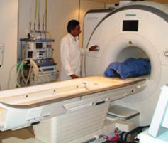
Skyra MRI with all clinical applications in the run since 28-Novemeber-2013.
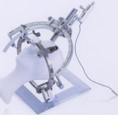
Inomed Riechert-Mundinger System, with three point
fixation is the most accurate system in the market. The microdrive and
its sensor gives feed back about the localization.
Inomed MER system
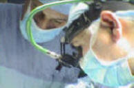
Leica HM500
The World's first and the only Headmounted Microscope.
Freedom combined with Outstanding Vision, but very bad video recording and
documentation.
After long years TRUMPF TruSystem 7500 is running with in the neurosuite at
Shmaisani hospital starting from 23-March-2014
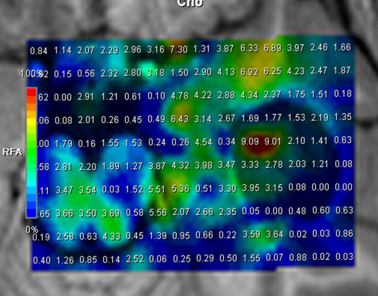
Choline distribution showing an active nidus medio-basal to the
mass.
Spectroscopy of the mass confirming glial nature of the mass.
Localization of the tumor according to data 22-July-2015 using
Inomed Planning sofware.
Notice: Not all operative activities
can be recorded due to lack of time.
Notice: Head injuries and very urgent surgeries are also
escaped from the plan .