Most of the site will reflect the ongoing surgical activity of Prof. Munir Elias MD., PhD. with brief slides and weekly activity. For reference to the academic and theoretical part, you are welcome to visit
neurosurgery.tv
Inomed Stockert Neuro N50. A versatile
RF lesion generator and stimulator for
countless applications and many uses
Multigen RF lesion generator .
29-MAY-2014 HUDA WAHEED AL-MASRI 61 YEARS TUMOR OF
THE OLFACTORY REGION WITH INTRADURAL GROWTH AND MASSIVE LEFT ETHMOID-SPHENOID
AND NASAL CAVITY.
Anamnesis
The patient came to the clinic 22-May-2014 after
performing biopsy through the left nostril
19-May-2014 at Jordan hospital with biopsy
result done 21-May-2014 telling that she has
poorly differentiated
sinonasal adnocarcinoma non-intestinal type.
The operation was done by CT-scan data done
14-May-2014 for the sinuses and was operated as
for polyp.
On examination; The patient has bilateral
anosmia for unknown period of time. She still
has bloody discharge from the nose. She is
neurologically free aside of anosmia. The
patient is right handed.
The patient was sent for new CT-scan of the
skull base and MRI of the brain and nasal
cavities with contrast with MRA of the brain and
carotids. She was advised to perform the
investigations after the clearing of the nasal
discharges. MRI and CT-scan done 26-May-2014
showing a tumor originating from the olfactory
bulbs invading the intradural space destroying
the planum sphenoidale and extending down to the
ethmoid cavities and left side of the sphenoid
sinus and the left maxillary cavity. It was
occluding the left nostril.
Bifrontal subfrontal craniotomy with reflection
of the bone flap to the left. Surgicele was
applied as usual over the superior sagittal
sinus as usual to avoid bleeding. The anterior
edge of the bone flap was created abutting the
anterior fossa plan, violating during that the
frontal sinuses. The tumor was fulfilling the
left part of the frontal sinus. It was sent to
fresh frozen biopsy, which gave the result of
malignant adenosarcoma. The tumor was violate
fleshy, friable. The dura was opened parallel to
the anterior edge of the bone defect. The
intradural tumor was totally resected. It was
completely destroying the left olfactory bulb
but the right one was anatomically intact and
was preserved. It was possible to close the dura
without applying a dural graft with preservation
of the right olfactory bulb. Resection of the
tumor in the nasal cavity was undertaken and the
tumor was completely destroying the
medio-inferior wall of the left orbit. The
orbital structures were preserved. So as to gain
more visual control, it was necessary to create
a small bony miniflap at the nasal bone to see
the tumor residing directly under the anterior
fossa. The tumor was completely destroying
the nasal septum, for what it was removed with
tumor. Drilling of the anterior wall of the
sphenoid sinus, which was also tumorous in
inspection. All the visible parts of the tumor
were resected. The dura was closed and the bone
flap reflected back to place and three separate
stitches were applied to the and bandaging of
the head was done. The patient was sent for MRI,
which showed a small residual of the tumor near
the exit of the left nostril and mild subdural
hematoma over the right convexity. The wound was
reopened as anew and seeking for the cause of
the right subdural hematoma was identified after
removing the surgicele. There was dural tear at
the posterior edge of bone defect in the right
side, which was reaching the lateral wall of the
superior sagittal sinus. The hematoma was
evacuated and the sinus tear was repaired and
the dura was water-tightly closed. The small
bone flap was returned to place and fixed and
the bifrontal flap was returned to place after
applying a piece of muscle to the bone defect at
the left substantia crebrosa with glue in both
side to keep in in place. Routine closure of the
wound. Before weaning the patient, the piece
near the exit of the left nostril, was removed
using specula through the nostril.
Smooth postoperative
recovery. The patient was sent to the ICU for 24
hour observation.
Comments
The patient has an aggressive tumor that
destroying the bony elements and growing intradural.
The histologic results were sent to 2
separate doctors with promise to perform full investigations
to have the final diagnosis without mistakes.
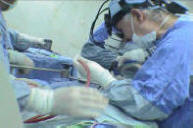
Intraoperative MRI seems to be mandatory
in cranial and such skull base surgery. Here the remnant at
the most inferior part of the nasal cavity was seen and
removed accordingly and the right sided subdural hematoma
was seen and the tear of the right side of the SSS was
repaired and the hematoma evacuated. The MRI control took
less than 10 min, but saved several days and weeks of the
possible complications.
Histologic results
The first result was that the neoplastic
cells are: EMA +, NSE focal +, S100 focal +, Chromogranin
rare positive cells, Synaptophysin focal+, Pan CK =, MNFCK
+, GFAP -, TTF1 -, MELAN A -, HMB 45 -, LCA -, CD99 -,
Vimentin -, MPO -. Sino-nasal tumor with intracranial
extension: High grade undifferentiated malignant tumor,
consistent with malignant neuroendocrine tumor ( Dr. Fayez
Hajjiri).
Anaplastic tumor of undetermined
histogenesis (Dr. Salah Al-Jitawi).
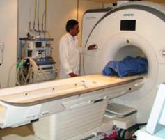
Skyra MRI with all clinical applications in the run since 28-Novemeber-2013.
Leica HM500
The World's first and the only Headmounted Microscope.
Freedom combined with Outstanding Vision, but very bad video recording and
documentation.
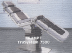
After long years TRUMPF TruSystem 7500 is running with in the neurosuite at
Shmaisani hospital starting from 23-March-2014
Notice: Not all operative activities
can be recorded due to lack of time.
Notice: Head injuries and very urgent surgeries are also
escaped from the plan .