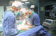
Most of the site will reflect the ongoing surgical activity of Prof. Munir Elias MD., PhD. with brief slides and weekly activity. For reference to the academic and theoretical part, you are welcome to visit
neurosurgery.tv
Inomed Stockert Neuro N50. A versatile
RF lesion generator and stimulator for
countless applications and many uses
Multigen RF lesion generator .
10-JUNE-2013 MUHAMED FALAH HASAN 57 YEARS
SPONDYLOLISTHESIS L5-6 WITH LUMBAR CANAL STENOSIS L4-5 AND L5-S1.
Anamnesis
The patient came to the clinic 07-January-2008
with LBP and right sciatica after suffering RTA
1999 in Cyprus with fracture pelvis at that
time. He got exacerbation of the right sciatica
the last three months. He is using crutches
during this period. Lumbosacral X-ray done
05-October-2007 showed old wedging fracture of
L2 with spondylolisthesis L5-S1. On examination
at that time: SLRS was 45 degrees with pain in
the right with weak dorsi and planterflexion
right foot 4/5. The AJs were absent both
sides. He had scoliotic stance. CT-scan of the
brain done 08-January-2008 was normal and MRI
lumbar spine showed spondylolisthesis I degree
of L5-S1 with old fracture L2 of no
significance. The patient was treated
conservatively.
The patient then came 12-September-2009 and
11-September-2012 with bilateral sciatica and
investigations showed the same spondylolisthesis
and stenosis at L4-5. The patient was willing
for conservative treatment.
The patent then came 29-May-2013 after
travelling to Malta, complaining that during
travel, he got severe agonizing LBP with
bilateral sciatica. He is diabetic for 3 years
and cardiac cath done 3 years ago was normal.
On examination: The patient is limping with
exaggerated scoliotic stance.
SLRS was 70 degrees both sides. There is weak dorsiflexion
both feet 4/5. All deep reflexes are absent. The
patient was advised to perform radiologic
studies in case of not improving.
MRI
lumbar spine done 03-June-2013 showing the old
wedged fracture of L2 of no clinical
significance and spondylolisthesis II degree at
L5-S1 with severe stenosis L4-5, L5-S1. Dynamic
studies showed the spondylolisthesis with
bilateral isthmolysis L5-S1.
After skin and facial
incision, CSF came out before performing bone
dissection from the space between L4 and L5
spinous processii. Laminectomy L4 and L5. Foraminotomy L5
and S1 roots both sides. The dura was deformed
and there was a dural pouch which was extending
between the L4-5 space. It was looking like a
synovial cyst, but after dural dissection it
turned to be a dural envelope which was
preserved and was closed by 6 zero nylon. The L5
lamina and it lateral masses were fail and the
upper edge of the lamia was tearing the dural
sac, which caused the dural pouch. All these
elements were removed. Right sided discectomy of
L5-S1. A Novel TL TLIF cage 9x10x30 mm was
inserted to the L5-S1 disc space. A bone graft
was inserted in the disc space. Using C-arm and
Isobar TTL Module In 6.2x45 mm polyaxial
screws were inserted to L5 body and 6.2x40 mm
screws to S1 body. 2 bended rods 5.5x25 mm and
60 mm length Easys cross connector were used to
fuse the L5, S1 level with mild compression.
Bone graft was added lateral to the rods.
Routine closure of the wound.
Smooth postoperative recovery. The power of
both feet became normal.
Comments
The patient has lumbar canal stenosis with
spondylolisthesis. All components must be
corrected.
The patient has CSF coming from the field before
reaching the dura. This means that the patient
had severe trauma recently. The dural defect was
repaired accordingly.
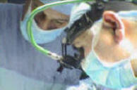
Leica HM500
The World's first and the only Headmounted Microscope.
Freedom combined with Outstanding Vision.
Notice: Not all operative activities
can be recorded due to lack of time.
Notice: Head injuries and very urgent surgeries are also
escaped from the plan .