Most of the site will reflect the ongoing surgical activity of Prof. Munir Elias MD., PhD. with brief slides and weekly activity. For reference to the academic and theoretical part, you are welcome to visit
neurosurgery.tv
Inomed Stockert Neuro N50. A versatile
RF lesion generator and stimulator for
countless applications and many uses
Multigen RF lesion generator .
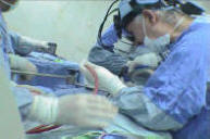
24-SEPTEMBER-2012 MUINEH MUHAMED SAADO 56 YEARS
CONDITION AFTER INCORRECT TRANSPEDICULAR FIXATION FOR SPONDYLOLISTHESIS L3-4.
Anamnesis
The patient came to the clinic 19-September-2012
complaining of difficult walking with severe
pain in the right knee for 18 years, which
increased after performed transpedicular
fixation in Syria 7 years ago. The patient is a
known diabetic and hypertensive for 1 year and
she underwent cath 11 years ago which was
normal. Exacerbation of the LBP and right
sciatica the last 6 months.
LSS X-ray done immediately after the surgery and
23-June-2012 showing spondylolisthesis L3-4 with
the right upper screw at L3 body out of the
bone. The left lower screw which was inserted to
the left L5 body was broken.
MRI lumbar spine done 23-June-2012 showing
spondylolisthesis L3-4 and bulge L4-5 and L5-S1.
On examination, the patient is limping with
exaggerated scoliotic stance with SLRS was 30
degrees both sides more painful in the right.
The KJ was absent in the left(?). There is weak
dorsi and planterflexion right foot 3/5. with
hypalgesia right L5 and S1 territories.
The old incision refreshed.
The screws were exposed with their knots. The
left lower screw was flail, so the right upper
screw. Both were removed with leaving the
fractured part inside the L5 body in place,
since it doesn't cause harm. Using Scientex
Alphatec Spine screws ISO bar TTL module 6.2x40
mm fixed screws were inserted to the right L3
pedicle and to the left L4 body. The previously
inserted screws were checked for looseness. They
were solid stable, for what they were left
untouched. The right side of the L3-4 of what to
be supposed to be the ligamentum flavum was
filled by the previous graft. It was drilled off
with high speed drill and foraminotomy of the
right L4 roots was achieved. Considering the
solid bony fusion of the L3-4 by the bone graft,
it was decided not to violate the L3-4 disc
space and the idea about inserting the TLIF was
withdrawn from the plan. The right L4 root was
severely compressed by the bony elements. Two
5.5 mm rods bended to accept the natural curve
of the spine were inserted and fusion of L3 and
L4 was achieved.
Routine closure of the wound. Dramatic
improvement of the right foot.
Please! wait for 3-5 min till the
video start to load. It depends upon the internet
connection.
Comments
The patient underwent
first surgery with the right upper screw
inserted lateral to the right pedicle. It could
irritate the the running L3 root and cause the
pain of the right knee which appeared
immediately after the first surgery. The second
possible cause is the severe compression of
right L4 root, which was decompressed by high
speed drill.
Leica HM500
The World's first and the only Headmounted Microscope.
Freedom combined with Outstanding Vision.
Notice: Not all operative activities
can be recorded due to lack of time.
Notice: Head injuries and very urgent surgeries are also
escaped from the plan .