Most of the site will reflect the ongoing surgical activity of Prof. Munir Elias MD., PhD. with brief slides and weekly activity. For reference to the academic and theoretical part, you are welcome to visit
neurosurgery.tv
Inomed Stockert Neuro N50. A versatile
RF lesion generator and stimulator for
countless applications and many uses
Multigen RF lesion generator .
09-AUGUST-2011 MUSA IBRAHEEM AHMAD KHALEEL 62
YEARS SUSPECTED INTRADURAL MASS IN THE CONUS MEDULLARIS WITH STENOSIS OF L2-3, 3-4 AND
L4-5.
Anamnesis
The
patient came to the clinic 31-July-2011
complaining of numbness both legs for one month
with progressing course, more the right with
unsteady gait and LBP. Right shoulder and neck
pain. The patient is a known diabetic under
treatment for 10 years, hypertensive for 10
years in Atakand, and his son noticed loss of
weight 12Kg the last 2 months.
MRI
cervical done 15-June-2011 showing
small PCD C2-3, 3-4, 4-5, 5-6 and C6-7. MRI
right knee showing tear meniscus with effusion.
On
examination: the patient is limping. Romberg
stance is stable, but cannot elevate the right
upper limb due to partial frozen right shoulder.
Both quadriceps femores are weak right
-4/5 left 4/5. Dorsiflexion right foot -4/5 and
left foot 3/5.
The
patient was sent for further investigations,
which revealed the presence of intradural mass
at the level L1 down to L4 in the conus
medullaris with stenosis at these levels. Dorsal
MRI was normal and MRI brain showed old
scattered lacunar infarctions both cerebral
hemispheres more the left side. Bone scan showed
only active site at the right shoulder. Right
shoulder MRI showed synovitis with partial tear
of the supraspinatus. CT-scan of the chest was
free.
It was explained to the
patient before the surgery, that decompression
is needed, but concerning the presence of tumor,
it was suggested that some patients have
abnormal cauda equina superlonga, that could
mimic a tumor when stenosis have place.
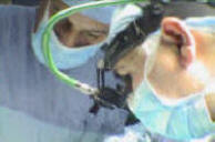
Decompressive laminectomy of
L2-3 and partial of L1 and L4. There was severe
elements of lumbar canal stenosis with absent
epidural fat. The patient was positioned with
head down to prevent CSF leak. The dura was
opened 30 mm along the most suspected tumor
location as reported by the radiologist. There
in no tumor, no seeders, no inflammatory
changes. The roots are tortuous, long and normal
looking. Inspection was proceeded entirely
between the roots and no data support the
presence of vascular malformation. The dura was
water-tightly closed with 6 zero nylon.
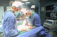
Routine
closure of the wound. Smooth postoperative
recovery .
Please! wait for 3-5 min till the
video start to load. It depends upon the internet
connection.
Comments
The patient is looking well and the patient with
metastasis usually looking toxic. It is rare to
see with localized intradural seeders without
the presence of primary tumor, which is usually
the posterior fossa in children.
Anatomical variations of the conus medullaris
could be variable, among them the super long
roots, which become tortuous and with presence
of stenosis mimic the presence of intradural
masses at the cauda equina and below.
For more information about the superlong roots
of the cauda equina press
here!
Notice: Not all operative activities
can be recorded due to lack of time.
Notice: Head injuries and very urgent surgeries are also
escaped from the plan .