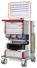
Inomed Stockert Neuro N50. A versatile
RF lesion generator and stimulator for
countless applications and many uses
Multigen RF lesion generator .
05-APRIL-2026 RABAA BADER AL-HNEYTI
59 YEARS EXTRUDED DISC L3-4 WITH LEFT FORAMINAL
AND EXTRAFORAMINAL OCCLUSION.
Anamnesis
The patient came to the clinic 08-September-2025 complaining of
agonizing left sciatica down to the heel left
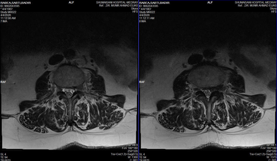
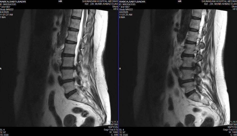
foot for 1 month with LBP. MRI lumbar spine done
27-August-2025 showing extruded disc L3-4 with
left up and extraforaminal migration.
On examination, the patient is not limping with
no scoliotic stances. SLRS
right side was 80
degrees with tightness and 60 degrees
in the left with pain. There is weak
dorsiflexion left foot 4/5. There is sensory
deficit left S1 root territory. Weak left
iliopsoas and left quadriceps 4/5. The patient
was given medication and advised for surgery,
but she disappeared. She is a known diabetic and
have arterial hypertension.
The patient then came urgently to the hospital
with agonizing pain for 2 days urging for
surgery 04-April-2026. MRI repeated showing the
same extrusion with more extraforaminal
extrusion and regression of the upward piece and cardio consultation was
asked.
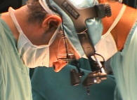
Prone position. The level of L3-4 was
identified and foraminotomy left L4 root was done.
Left sided L3-4 disc cleaning was done with
removal of the left far lateral migrating extrusion.
Using
MultiGen, bipolar stimulation of the left L4
root responded to 1.7 Volts. A bipolar pulsed
mode RF with 42 Celsius, 240 sec, 2 Hz and 20
msec duration to the left L4 root was
achieved using 2 bended catheters 10 mm exposed
length. Further bipolar stimulation of left L4
root responded to 1.2 Volts. The patient was put in Reverse
Trendelenburg position with Valsalva maneuver
and hyperventilation. No CSF leak. Routine closure of the
wound. The patient showed dramatic recovery. She was sent to the ward.
MultiGen
FOLLOW UP
The patient then came 11-April-2026 with
agonizing left sciatica and MRI done urgently
showing huge recurrence of the extrusion of L3-4
left side. The patient then urgently was
transferred to the operative room and the wound
was opened. There was still leaking fluid in the
area of the wound due to diabetic mellitus with
the extrusion was compressing the left L4 root.
Further drilling lateral left side and more
foraminotomy of the left L4 root. The extruded
swollen piece was removed and further cleaning
of the disc space was achieved. Routine closure
of the wound and intraoperative MRI was done
before weaning to the patient was done,
confirming disappearance of the extrusion.
There was no video recording because all
measures were taken urgently. Routine closure
and the sciatic a disappeared.
The patient was discharged 13-April-2026.
Comments
The extruded disc was occluding the left
L4 root and surgery will improve the related to the
extrusion problems.
This is the 297th case using the MultiGen. This procedure regained routine acceptance.
It became a usual part of the spine and peripheral nerves
surgery. Click here
for reference. The patient showed improvement of the motor
stimulation after BPRF and the sciatic pain disappeared and
regained almost normal power of the left foot.
With accumulation of data, it became
clear that the irritated nerve with aberrant currents
running in the C fibers up, not only causing no change or elevation of
the required voltage to achieve motor response, but they could cause the preoperative
weakness. Ablation of such currents results in facilitation
of the motor response and improvement of function with
disappearance of pain.
It is unclear why the roots have several
motor response with different patients, despite the fact
that the neurological status was the same and the anesthesia
protocol also the same.
It could be that the nerve is recovering
minute by minute after decompression and this can explain
why the motor conductivity is improving after the BPRF
application, which require 5 minute session in most cases.
After the 172d case, the elevation of
motor stimulation above 5 V was abandoned to avoid delayed
dural tear with subsequent CSF leak, which take place at the
contact at the lower electrode shaft with the dura below or
above the
level of the axilla.
Before doing motor stimulation in
peripheral nerve surgery with tourniquet. always release the
tourniquet before performing motor stimulation.
In this particular case the recurrence
was related to her diabetes mellitus which caused swelling
of the annulus fibrosis.
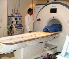
Skyra MRI with all clinical applications in the run since 28-Novemeber-2013.
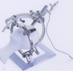
Inomed Riechert-Mundinger System, with three point
fixation is the most accurate system in the market. The microdrive and
its sensor gives feed back about the localization.
Inomed MER system
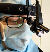
Leica HM500
The World's first and the only Head mounted Microscope.
Freedom combined with Outstanding Vision, but very bad video recording and
documentation.
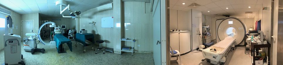
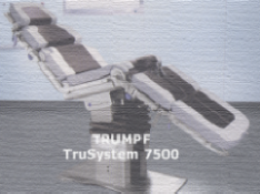
After long years TRUMPF TruSystem 7500 is running with in the neurosuite at
Shmaisani hospital starting from 23-March-2014
LooksCam II Xenosys in the run starting from 14-March-2021 with
SheerVision TTL x4 magnification.
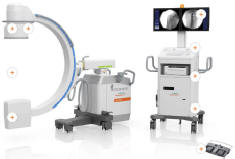
Cios-Spin flat panel in the run.
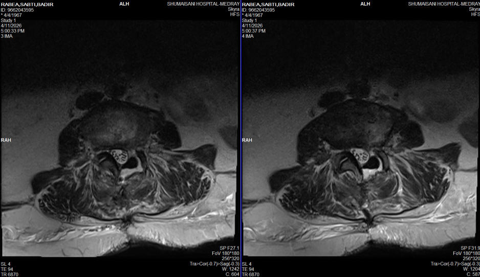
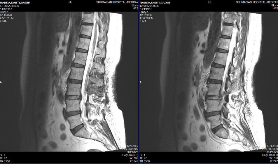
MRI done 11-April-2026 showing the recurrence axial view
MRI done 11-April-2026 showing the recurrence sagittal view
Intraoperative MRI confirming removal of the disc at L3-4, axial
view
Intraoperative MRI confirming removal of the disc at L3-4, saggital
view
Notice: Not all operative activities
can be recorded due to lack of time.
Notice: Head injuries and very urgent surgeries are also
escaped from the plan .