Most of the site will reflect the ongoing surgical activity of Prof. Munir Elias MD., PhD. with brief slides and weekly activity. For reference to the academic and theoretical part, you are welcome to visit
neurosurgery.tv
The
patient is an Iraqi child came to the clinic
22-April-2012. The parents notices a lump in the
middle of the lumbar area since birth.
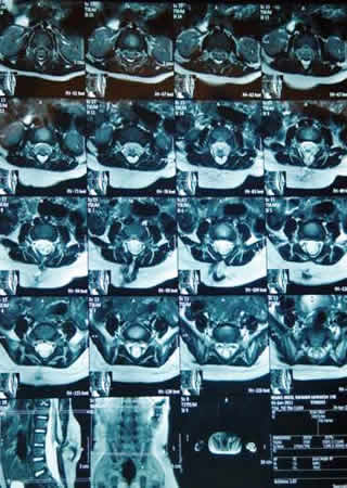
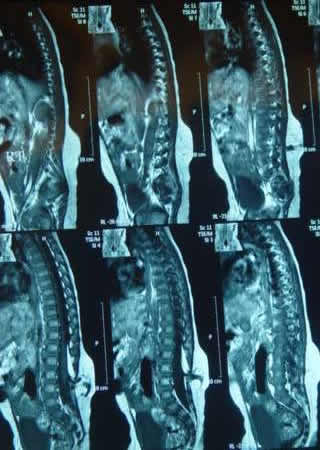
MRI of the
lumbar spine performed 23-October-2011 of bad
quality.
The
patient is neurologically free with a lump in
mid of the lumbar area with bony deformity more
prominent in the left side. The skin over the
lesion is intact but pulsating with CSF felt
under the skin. There is slight atrophy of the
right scapula in comparison with the right.
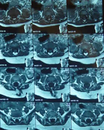
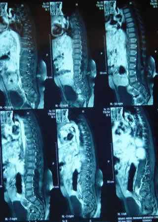
The
patient was sent to perform MRI of the brain and
the cervical spine and the lumbar area with
contrast. It was done 24-April-2012 confirming
the presence of small lipoma and tethering of
the spinal cord to the neck of the sac. MRI of
the brain and cervical spine were normal.
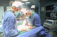
Skeletonization of L3, L4 and
L5 was done. The bony lesion which was not in
continuity with the spinal column was removed.
Partial laminectomy of L3 was done to expose the
dura and downward dissection was carried out
until the sac was identified and dissected off.
The CSF is coming free from the dural defect.
The defect was inspected and the lipoma was
still tethering the cord above the defect, for
what, complete laminectomy of L3 was done and
the dural defect was extended upward around 12
mm. The lipoma was identified and partial
resection down to the boundaries of the neural
structures to preserve them and to have complete
untethering of the spinal cord. All the
tethering elements were sharply dissected
including the arachnoid layers. Water tight
closure of the dura.
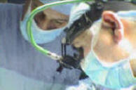
Routine
closure of the wound. Smooth postoperative
recovery.
Please! wait for 3-5 min till the
video start to load. It depends upon the internet
connection.
Comments
The aim of the
surgery is to untether the spinal cord and to
yield the cushioning effect of the dural defect
due to mechanical irritation.
Notice: Not all operative activities
can be recorded due to lack of time.
Notice: Head injuries and very urgent surgeries are also
escaped from the plan .