|
The patient started to complain of severe headache with ataxia and
blurred vision the last 45 days with progressive course. The patient
mentioned constant fall to the right and nausea with vomiting
attacks. MRI performed showing a huge well circumscribed mass in the
left cerebellum. On examination, aside to the cerebellar signs
there were no motor or sensory deficit. The patient was operated in
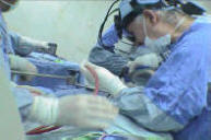
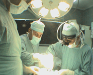
the setting position and osteoplastic craniotomy over the left
cerebellar convexity, radical removal of the mass was achieved and
the frozen section demonstrated a malignant nature of the tumor
without giving more details about the exact nature of the tumor.
The tumor was well defined with low vascularity and had good
cleavage. It was totally removed. Smooth postoperative recovery.
The final histologic result was that of medulloblastoma. For
theoretical data concerning medulloblastomas
click here!
Control CT-scan performed the same day of the operation and
the morning of the second day were acceptable. The second night
08-August at 3.15 a.m. the patient started to complain of headache
followed within seconds with difficulty in breathing and sudden
apnea. She was put immediately in the ventilator . During that, the
right pupil was fully dilated. Emergency CT-scan was performed and
the morphologic picture as the same as before. During CT-scan
investigation both pupils became fully dilated and it was
decided to urgently take the patient in the clinical basis to
explore the area.
The bone flap removed and the bone defect widened and the dura
was opened. Removal of the laceration and edematous cerebellar
tissues was undertaken and the CSF became to drain from the cisterna
magna and the ponto-cerebellar angle after what the brain regained
pulsation and the CSF start to flow freely. 35 minutes passed since
the progression of the bilateral full dilated non-reactive pupils,
after what the pupils became normal with sluggish reaction to light.
The dural defect was left and lyodura was placed over the pulsating
brain tissues. The patient was kept in ventilator.
The patient after 2 days in ventilator another time progressed
dilated right pupil, for what repeat CT-scan was performed and
showed swelling both cerebellar hemispheres and the supratentorial
compartment. She was taken to the operating room and a wide bone
flap was removed over both cerebellar hemispheres and both occipital
lobes were exposed. Transverse incision was made over both
cerebellar hemispheres with resection of the falx cerebelli. The
right cerebellar hemisphere bulged with pressure and start to
pulsate and attempt to put canula to the posterior horns failed due
to slit ventricles. The wound was closed superficially and the
pupils regained normal position after 20-30 min. The patient, then
continued to deteriorate despite aggressive measures and polyuria
took place the morning of 12-August-2006 and diabetes
incipidus was established and minrin started.
The patient progressed mild dilatation of the right pupil
14-August, and she died the morning of 15-August-2006.
|