Most of the site will reflect the ongoing surgical activity of Prof. Munir Elias MD., PhD. with brief slides and weekly activity. For reference to the academic and theoretical part, you are welcome to visit
neurosurgery.tv
Inomed Stockert Neuro N50. A versatile
RF lesion generator and stimulator for
countless applications and many uses
Multigen RF lesion generator .
10-DECEMBER-2013 SHAIMA FALAH HASAN TB OF THE
QUADRIGEMINAL AREA AND TENTORIUM AND PACHYMENINGITIS OF THE DORSAL SPINE FROM D2
DOWN TO D12 WITH SEVERE COMPRESSION OF THE SPINAL CORD FROM D8-D11.
Anamnesis
The patient came to the clinic 07-December-2013
in wheelchair complaining of severe weak upper
and lower limbs and inability to walk the last
three weeks. During last months of pregnancy got
fever in June and July this year with numbness
of the left upper limb and blindness left eye.
Delivery was 10-August-2013. 10 days later she
deteriorated with headache and pain four limbs.
The patient was investigated and treated for tb.
The last three weeks she became bedridden
and in wheelchair with lost sensation for
urination and defecation. MRI of the brain done
01-September-2013 showing lesion in the pineal
area. MRI cervical and dorsal done
18-September-2013 of bad quality not
informative. MRI dorsal repeated
28-November-2013 showing pachymeningitis of the
dorsal spine.
On examination is in wheelchair, vision left eye
improved, but still having mild left abducens
paresis. There is weak extensors both hands 4/5,
triceps both upper limbs 4/5. There is profound
hypalgesia below D3 both sides. The right
quadriceps femoris 4/5, left 3/5. Knee adductors
and adductors 3/5 weaker in the left. Dorsi and
planterflexion both feet 3/5. Babinski positive
both sides. Loss of urination and defecation
control.
MRI of the brain and whole spine with contrast
and spectroscopy was done using Skyra magnetom
07-December-2013. The mass occupying the whole
quadrigeminal cistern regressed, but there are
scattered gliotic changes of the left pulvinar
area and small spots at the edges of the
tentorium without mass effect. There is
pachymeningitis starting from D2 down to D12
with severe compression of the spinal cord at D8
down to D12. Spectroscopy ruled out malignancy.
The patient was admitted 08-December-2013 and
one unit of blood was given and infection
specialist started treatment.
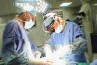
Inomed ISIS IOM was attached
with dorsal spine tumor protocol. Using C-arm, laminectomy of
D8 and D9. There is no epidural fat and no
pathologic material out of the dura or the bone
structures. The dura was opened and the rubbery
like material filling the subarachnoid spaces
compressing the spinal cord circumferentially.
The layer which was as a carpet with
granulomatous lesions was diffusely adherent to
the spinal cord. Using sharp dissection it was
possible to dissect part of the posterior parts
of the pathologic material and it was sent for
fresh frozen biopsy. It gave the answer of
inflammatory process of unknown character. The
granulomatous nodules were decompressed, so as
to prevent surgical damage to the spinal cord.
Feeding tube No 6 was inserted up to 25 cm and
CSF came out and sent for all investigations.
There was also debris of the pathologic material
which was sent with the excised granulomas for
histologic studies. Using Omipaque
myelography was done up and down to see the
limits of severe compression. It was decided to
extend the dural incision 1 vertebra above and
one vertebra down. The dural defect was closed
with lyodura to achieve good volume to the
spinal cord and surrounding pathologic ring.
Water-tight closure of the lyodura with nylon 4
zero. Routine closure of the wound.
Smooth postoperative
recovery. The power of the upper limbs became
normal and slight improvement of the power of
both lower limbs.
Comments
This case is very rare. The pathologic material
was rubbery strangulating and compressing the
spinal cord. Surgical decompression at all
layers was mandatory to provide good circulation
to the affected spinal cord.
The pathological process was constricting the
spinal cord as a band. It was necessary to
release this band.
Leica HM500
The World's first and the only Headmounted Microscope.
Freedom combined with Outstanding Vision.
Notice: Not all operative activities
can be recorded due to lack of time.
Notice: Head injuries and very urgent surgeries are also
escaped from the plan .