|
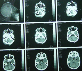
The patient 5 years old with right
sixth nerve paresis since childhood got for several weeks unsteady
gait with progressing headache and blurred vision. MRI preformed
12-November-2005 showed a huge vascular mass in the right cerebellar
hemisphere compressing the brain stem and causing hydrocephalus.
Romberg was unstable swaying to all directions. Otherwise, except
for the right 6th nerve paresis was intact.
The patient was operated in the
sitting position with osteoplastic suboccipital craniotomy of my
personal modification, with pedicle to ligamentous structures to C1
and was reflected inferiorly. V-shaped incision of the dura. The
vascular lesion was attacked from several direction, trying during
that to preserve the most tiny normal vascular architecture. The
main feeders were arising from the right PICA. It had no
communication with the transverse or sigmoid sinus as was
anticipated. The vascular mass was resected totally. The
cerebellum was hanging free with good cardio-pulmonary pulsation.
Water-tight closure of the dura and the flap returned back.
Postoperative period was smooth
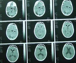
and the patient showed no complications. Postoperative CT-scan
performed the next day confirm the radical resection of the mass and
absence of any complications.
For theoretical information about
cavernous heamangiomas, click
here!


|