Clinical Presentation
and Diagnosis
Glossopharyngeal neuralgia was defined as a clinical entity by Harris in
1921. It is characterized by paroxysms of pain in the sensory
distribution of the ninth cranial nerve. Except for the location of the
pain and the sensory stimuli which induce it. the attacks are identical
to those of trigeminal tic douloureux. The incidence of glossopharyngeal
neuralgia is one-seventieth that of trigeminal neuralgia' The typical
pain is a severe lancinating repetitive series of electric-like stabs in
the region of the tonsil or posterior one-third of the tongue
unilaterally. Bilateral glossopharyngeal neuralgia was never encountered
in Dandy's series or in Mayo Clinic experience. The pain may either
radiate to or originate in the ear.
The sensory stimulus which induces the
pain is swallowing and during severe attacks the patient may sit
motionless. head flexed forward, allowing saliva to freely drool
from the mouth. Although remissions may occur for months or years.
spontaneous cure has never been reported. Associated with attacks of
glossoşpharyngeal neuralgia have been cardiac arrest, syncope, and
seizures. Cardiac arrest occurring during an attack of glossopharyngeal
neuralgia has been attributed to hypersensitivity of the dorsal motor
nucleus of the vagus caused by repetitive afferent impulses from the
pharynx. tragus and ear. These impulses. occurring during an attack of
neuralgia or secondary to surgical manipulation. may also be conveyed to
the dorsal motor nucleus via collaterals from the nucleus solitarius of
the ninth nerve causing asystole. Carotid sinus syncope and seizures
occur by this latter mechanism. causing reduced cardiac output and
cerebral hypoxia.
Almost
all cases of glossopharyngeal neuralgia are idiopathic. although a
certain number have been ascribed to entities such as ossification of
the stylohyoid ligament nasopharyngeal and cerebellopontine angle
tumors, an atheromatous vertebral artery causing compression of the
ninth nerve, anomalous vascular lesions, tortuous vertebral and
basilar arteries. and arterial loops. Historically. Weisenburg in
1910 first described glossopharyngeal neuralgia in a patient harboring
an extra-axial posterior fossa tumor.
When ear
pain is predominant. Svien et al. pointed out that five separate nerves
must be considered: the tympanic branches of the glossopharyngeal. the
auriculotemporal branch of the trigeminal. the nervus intermedius. the
auricular branch of the vagus. and the upper fibers of the
cervical nerves. Robson and Bonica emphasized the overlapping of
sensory neuralgias and the rare diagnostic dilemmas posed by pain at junctional sites between the pharynx and
ear. Cocainization of the
pharynx alleviates the ninth nerve component of pain. and cocainization
of the pyriform fossa relieves neuralgia of the superior laryngeal
branch of the vagus. By blocking the foramen ovale with bupivacaine. it
is possible to determine the component of pain due to the third
division of the trigeminal nerve. Tetracaine block of the jugular
foramen will block all afferent impulses via the ninth and tenth nerves
and help to sort out the rare pain mediated only by the nervus
intermedius component of the seventh cranial nerve.
Treatment
Adson reported temporary relief in two
patients in whom he sectioned the glossopharyngeal nerve extracranially
but encountered great technical difficulty because of its close
proximity to the vagus nerve and jugular bulb. He later
performed cadaver dissections and described a suboccipital
approach to intracranial ninth nerve sectioning, but in 1927 Dandy
reported the first intracranial ninth nerve section for the relief of glossopharyngeal neuralgia. Dandy, Fay and others in earlier
report, stressed the need for sectioning the upper one-sixth to one-eighth
of the vagus rootlets to prevent recurrence of pain. It has later been reemphasized
that the superior laryngeal branch of the vagus and the
tympanic plexus component of the vagus may be not infrequent
concomitant of glossopharyngeal neuralgia.
Since, section of the ninth cranial nerve
and the upper rootlet of the tenth leaves the patient with no
discernible sequelae, it would seem unwise to subject the patient
needlessly to the possibility of recurrence by microvascular
decompression of the ninth nerve alone.
The
percutaneous radiofrequency lesions which have been suggested in the
past seem fraught with an inordinate risk of injury to the tenth
cranial nerve. Tew described percutaneous radiofrequency procedures
carried out in nine patients with pharyngeal pain secondary to neoplasms of the head and neck and
in two patients with idiopathic
glossopharyngeal neuralgia. Excellent relief was obtained from the
pain of malignant disease: however, vocal cord paralysis occurred after
rhizotomy in both patients treated for idiopathic pain and aspiration pneumonia in one.
When malignant disease has already altered the swallowing mechanism. radiofrequency lesions seem tenable: however,
when
tenth nerve preservation is mandatory, the procedure is undesirable.
Operative Approach
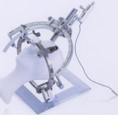
The patient is placed in the upright
sitting position in the pinion headrest. with the head acutely flexed
and rotated to the side of the glossopharyngeal neuralgia. A central right atrial catheter for monitoring and aspiration of
possible air emboli is placed before the patient is placed in the
sitting position. Either an S-shaped or a hockey stick-shaped incision
is made over the ipsilateral occipital bone. The 4-cm craniectomy is
done inferiorly to incorporate the portion of the occipital bone
which lies directly adjacent to the foramen magnum and which is oriented
in a tranverse plane directly above the lamina of the first cervical
segment. The dura is opened in a cruciate fashion and tacked back over
the edge of the craniectomy. The cerebellar hemisphere is elevated to
expose the arachnoid of the cisterna magna. which is opened. allowing
the egress of spinal fluid and relaxation of the cerebellum. By
identifying the sigmoid sinus as it traverses the posterior fossa floor.
the surgeon can achieve precise retractor position for identification of
the jugular foramen. Once the self-retaining retractor has been fixed in
position. illumination by the overhead surgical lights and operating
loupes is usually sufficient. The operating microscope may enhance
visualization at this point. The ninth cranial nerve in the jugular
foramen is always separated by the dural septum from the tenth and
eleventh cranial nerves and jugular vein.
The ninth nerve and upper one-sixth to
one-eighth of the filaments of the tenth nerve are sectioned with the
aid of a black spatula or blunt hook and bipolar coagulation. The dural
opening may then be closed by any of various methods. either with or without the aid of a fascia lata or homologous
dural graft.
Intra- and Postoperative
Considerations
While
sensation is diminished over the pharynx and the gag reflex is abolished
on the side of the divided nerve and while discrete neurological
testing reveals absence of taste over the ipsilateral posterior one-third of the
tongue, it is seldom to note more than a transient disturbance in
swallowing.
Intraoperative cardiovascular
complications during suboccipital craniectomy for glossopharyngeal nerve
section and decompression have been well documented. Jannetta
described one case of microvascular decompression which was followed by
a hypertensive crisis and a fatal intracerebellar hemorrhage. In
another case he described significant hypertension lasting for 1 week postoperatively.
Nagashima et al. summarized the
physiologic intraoperative changes in a case report. During an
intracranial procedure for sectioning the ninth cranial nerve. they
noted that an extrasystole occurred each time the vagus nerve was
touched and that acute hypotension followed. which responded to
atropine. During the hypotensive episodes there was electrocardiographic
evidence of a right bundle branch block secondary to coronary artery
insufficiency and myocardial ischemia with progressive ST segment
depression.
With section of the ninth and upper
rootlets of the tenth cranial nerves. auricular flutter. tachycardia.
hypertension. ectopic ventricular contractions and cardiac arrhythmias
have been noted. Medical
therapy with phenytoin or carbamazepine is of considerable use in some
patients to afford temporary and short-term pain relief. Although there
are obvious operative risks. as previously mentioned. the operative
results with sectioning of the ninth cranial nerve and a portion of the
tenth cranial nerve are uniformly excellent for total pain relief from
glossopharyngeal neuralgia. Unless there are unusual circumstances.
this is not only the procedure of choice.
